Calcium in PDB 5fk0: Yeast Delta-Cop-I Mu-Homology Domain
Protein crystallography data
The structure of Yeast Delta-Cop-I Mu-Homology Domain, PDB code: 5fk0
was solved by
R.J.Suckling,
P.R.Evans,
D.J.Owen,
with X-Ray Crystallography technique. A brief refinement statistics is given in the table below:
| Resolution Low / High (Å) | 48.41 / 3.00 |
| Space group | P 21 21 21 |
| Cell size a, b, c (Å), α, β, γ (°) | 86.900, 148.630, 222.720, 90.00, 90.00, 90.00 |
| R / Rfree (%) | 19.5 / 23.33 |
Calcium Binding Sites:
The binding sites of Calcium atom in the Yeast Delta-Cop-I Mu-Homology Domain
(pdb code 5fk0). This binding sites where shown within
5.0 Angstroms radius around Calcium atom.
In total 2 binding sites of Calcium where determined in the Yeast Delta-Cop-I Mu-Homology Domain, PDB code: 5fk0:
Jump to Calcium binding site number: 1; 2;
In total 2 binding sites of Calcium where determined in the Yeast Delta-Cop-I Mu-Homology Domain, PDB code: 5fk0:
Jump to Calcium binding site number: 1; 2;
Calcium binding site 1 out of 2 in 5fk0
Go back to
Calcium binding site 1 out
of 2 in the Yeast Delta-Cop-I Mu-Homology Domain
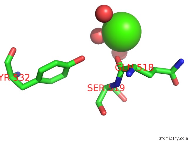
Mono view
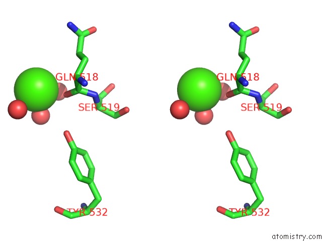
Stereo pair view
Mono view
Stereo pair view
A full contact list of Calcium with other atoms in the Ca binding
site number 1 of Yeast Delta-Cop-I Mu-Homology Domain within 5.0Å range:
|
Calcium binding site 2 out of 2 in 5fk0
Go back to
Calcium binding site 2 out
of 2 in the Yeast Delta-Cop-I Mu-Homology Domain
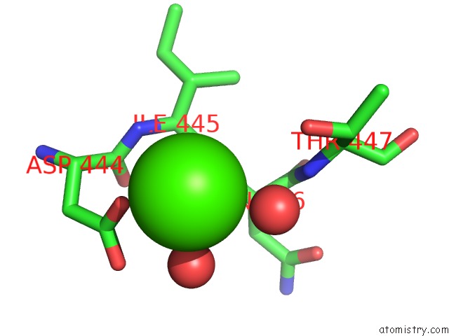
Mono view
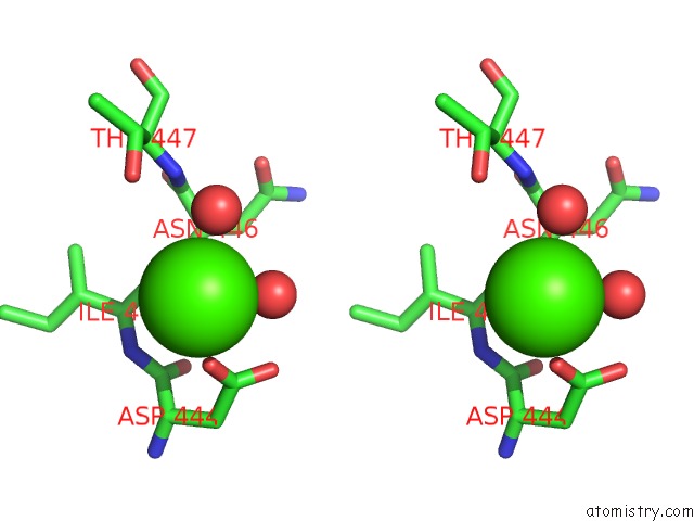
Stereo pair view
Mono view
Stereo pair view
A full contact list of Calcium with other atoms in the Ca binding
site number 2 of Yeast Delta-Cop-I Mu-Homology Domain within 5.0Å range:
|
Reference:
R.J.Suckling,
P.P.Poon,
S.M.Travis,
I.V.Majoul,
F.M.Hughson,
P.R.Evans,
R.Duden,
D.J.Owen.
Structural Basis For the Binding of Tryptophan-Based Motifs By Delta-Cop. Proc.Natl.Acad.Sci.Usa V. 112 14242 2015.
ISSN: ISSN 0027-8424
PubMed: 26578768
DOI: 10.1073/PNAS.1506186112
Page generated: Sun Jul 14 19:16:55 2024
ISSN: ISSN 0027-8424
PubMed: 26578768
DOI: 10.1073/PNAS.1506186112
Last articles
Zn in 9J0NZn in 9J0O
Zn in 9J0P
Zn in 9FJX
Zn in 9EKB
Zn in 9C0F
Zn in 9CAH
Zn in 9CH0
Zn in 9CH3
Zn in 9CH1