Calcium in PDB 7msy: Structure of CALU17 From the Calicheamicin Biosynthesis Pathway of Micromonospora Echinospora
Protein crystallography data
The structure of Structure of CALU17 From the Calicheamicin Biosynthesis Pathway of Micromonospora Echinospora, PDB code: 7msy
was solved by
A.J.Kosgei,
M.D.Miller,
W.Xu,
S.G.Van Lanen,
J.S.Thorson,
G.N.Phillips Jr.,
with X-Ray Crystallography technique. A brief refinement statistics is given in the table below:
| Resolution Low / High (Å) | 48.23 / 2.21 |
| Space group | P 43 21 2 |
| Cell size a, b, c (Å), α, β, γ (°) | 53.465, 53.465, 223.468, 90, 90, 90 |
| R / Rfree (%) | 18.4 / 23.6 |
Other elements in 7msy:
The structure of Structure of CALU17 From the Calicheamicin Biosynthesis Pathway of Micromonospora Echinospora also contains other interesting chemical elements:
| Chlorine | (Cl) | 2 atoms |
| Magnesium | (Mg) | 1 atom |
Calcium Binding Sites:
The binding sites of Calcium atom in the Structure of CALU17 From the Calicheamicin Biosynthesis Pathway of Micromonospora Echinospora
(pdb code 7msy). This binding sites where shown within
5.0 Angstroms radius around Calcium atom.
In total only one binding site of Calcium was determined in the Structure of CALU17 From the Calicheamicin Biosynthesis Pathway of Micromonospora Echinospora, PDB code: 7msy:
In total only one binding site of Calcium was determined in the Structure of CALU17 From the Calicheamicin Biosynthesis Pathway of Micromonospora Echinospora, PDB code: 7msy:
Calcium binding site 1 out of 1 in 7msy
Go back to
Calcium binding site 1 out
of 1 in the Structure of CALU17 From the Calicheamicin Biosynthesis Pathway of Micromonospora Echinospora
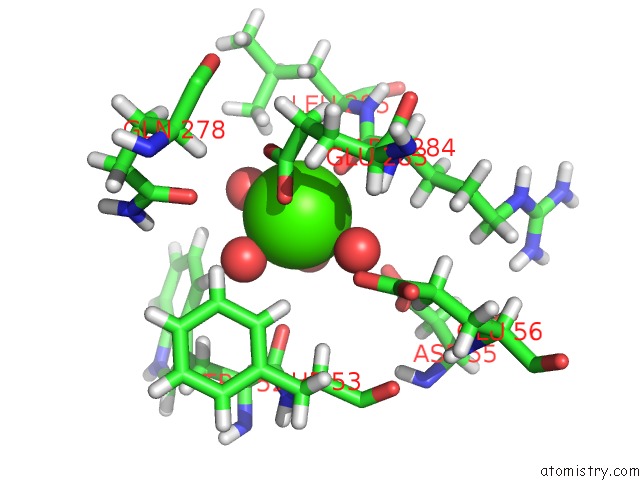
Mono view
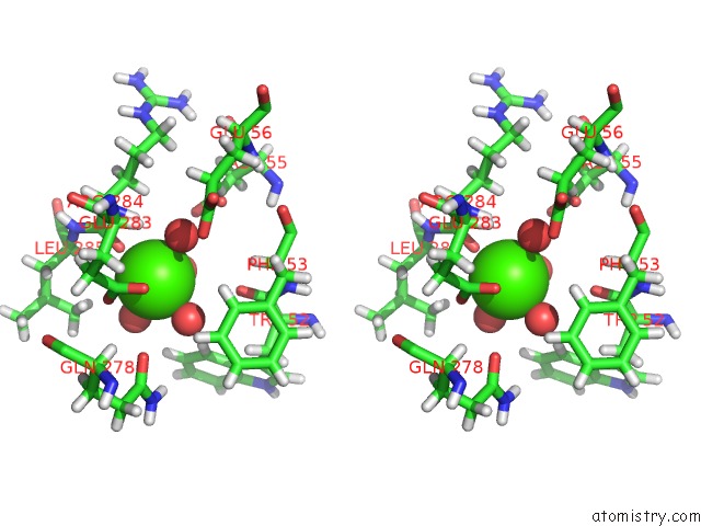
Stereo pair view
Mono view
Stereo pair view
A full contact list of Calcium with other atoms in the Ca binding
site number 1 of Structure of CALU17 From the Calicheamicin Biosynthesis Pathway of Micromonospora Echinospora within 5.0Å range:
|
Reference:
A.J.Kosgei,
M.D.Miller,
W.Xu,
S.G.Van Lanen,
J.S.Thorson,
G.N.Phillips Jr..
Structure of Dynf From the Dynemicin Biosynthesis Pathway of Micromonospora Chersina To Be Published.
Page generated: Fri Jul 19 02:09:02 2024
Last articles
K in 9G9VK in 9DTR
K in 9C46
K in 9G9W
K in 9G9X
K in 9ESI
K in 9ESH
K in 8ZEX
K in 8VAV
K in 8VAZ