Calcium in PDB 7ob0: Structure of Rslov D2 Variant
Protein crystallography data
The structure of Structure of Rslov D2 Variant, PDB code: 7ob0
was solved by
A.Moeglich,
T.G.A.Krafft,
M.Weyand,
with X-Ray Crystallography technique. A brief refinement statistics is given in the table below:
| Resolution Low / High (Å) | 42.93 / 1.90 |
| Space group | P 21 21 21 |
| Cell size a, b, c (Å), α, β, γ (°) | 58.057, 77.168, 155.002, 90, 90, 90 |
| R / Rfree (%) | 17.6 / 21.8 |
Other elements in 7ob0:
The structure of Structure of Rslov D2 Variant also contains other interesting chemical elements:
| Sodium | (Na) | 1 atom |
| Magnesium | (Mg) | 1 atom |
Calcium Binding Sites:
The binding sites of Calcium atom in the Structure of Rslov D2 Variant
(pdb code 7ob0). This binding sites where shown within
5.0 Angstroms radius around Calcium atom.
In total 2 binding sites of Calcium where determined in the Structure of Rslov D2 Variant, PDB code: 7ob0:
Jump to Calcium binding site number: 1; 2;
In total 2 binding sites of Calcium where determined in the Structure of Rslov D2 Variant, PDB code: 7ob0:
Jump to Calcium binding site number: 1; 2;
Calcium binding site 1 out of 2 in 7ob0
Go back to
Calcium binding site 1 out
of 2 in the Structure of Rslov D2 Variant
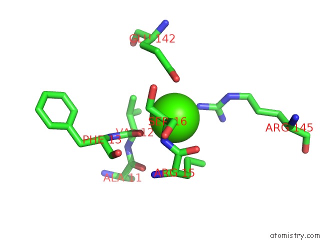
Mono view
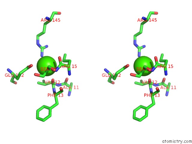
Stereo pair view
Mono view
Stereo pair view
A full contact list of Calcium with other atoms in the Ca binding
site number 1 of Structure of Rslov D2 Variant within 5.0Å range:
|
Calcium binding site 2 out of 2 in 7ob0
Go back to
Calcium binding site 2 out
of 2 in the Structure of Rslov D2 Variant
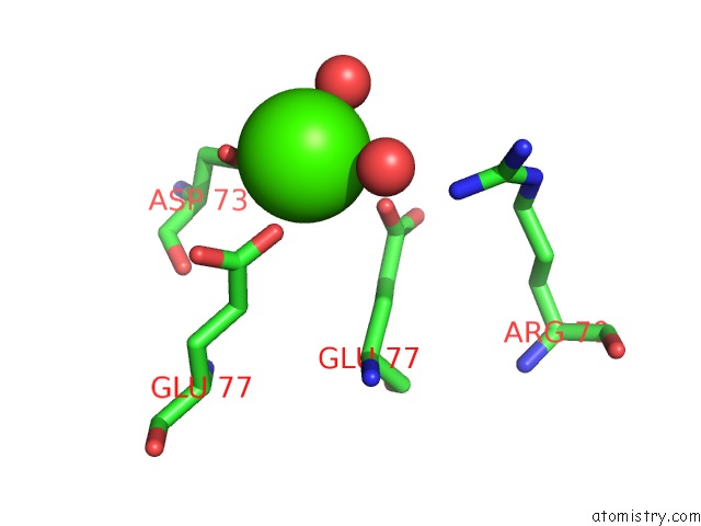
Mono view
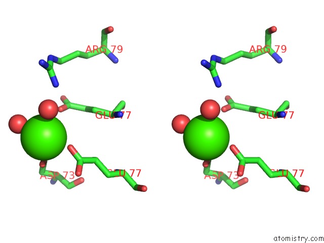
Stereo pair view
Mono view
Stereo pair view
A full contact list of Calcium with other atoms in the Ca binding
site number 2 of Structure of Rslov D2 Variant within 5.0Å range:
|
Reference:
J.Dietler,
R.Schubert,
T.G A Krafft,
S.Meiler,
S.Kainrath,
F.Richter,
K.Schweimer,
M.Weyand,
H.Janovjak,
A.Moglich.
A Light-Oxygen-Voltage Receptor Integrates Light and Temperature. J.Mol.Biol. 67107 2021.
ISSN: ESSN 1089-8638
PubMed: 34146595
DOI: 10.1016/J.JMB.2021.167107
Page generated: Fri Jul 19 02:28:48 2024
ISSN: ESSN 1089-8638
PubMed: 34146595
DOI: 10.1016/J.JMB.2021.167107
Last articles
Zn in 9J0NZn in 9J0O
Zn in 9J0P
Zn in 9FJX
Zn in 9EKB
Zn in 9C0F
Zn in 9CAH
Zn in 9CH0
Zn in 9CH3
Zn in 9CH1