Calcium in PDB 7oih: Glycosylation in the Crystal Structure of Neutrophil Myeloperoxidase
Enzymatic activity of Glycosylation in the Crystal Structure of Neutrophil Myeloperoxidase
All present enzymatic activity of Glycosylation in the Crystal Structure of Neutrophil Myeloperoxidase:
1.11.2.2;
1.11.2.2;
Protein crystallography data
The structure of Glycosylation in the Crystal Structure of Neutrophil Myeloperoxidase, PDB code: 7oih
was solved by
L.Krawczyk,
S.Semwal,
J.Bouckaert,
with X-Ray Crystallography technique. A brief refinement statistics is given in the table below:
| Resolution Low / High (Å) | 38.99 / 2.60 |
| Space group | C 1 2 1 |
| Cell size a, b, c (Å), α, β, γ (°) | 155.91, 144.634, 236.454, 90, 91.53, 90 |
| R / Rfree (%) | 17.9 / 22 |
Other elements in 7oih:
The structure of Glycosylation in the Crystal Structure of Neutrophil Myeloperoxidase also contains other interesting chemical elements:
| Iron | (Fe) | 8 atoms |
| Chlorine | (Cl) | 36 atoms |
| Fluorine | (F) | 4 atoms |
Calcium Binding Sites:
The binding sites of Calcium atom in the Glycosylation in the Crystal Structure of Neutrophil Myeloperoxidase
(pdb code 7oih). This binding sites where shown within
5.0 Angstroms radius around Calcium atom.
In total 8 binding sites of Calcium where determined in the Glycosylation in the Crystal Structure of Neutrophil Myeloperoxidase, PDB code: 7oih:
Jump to Calcium binding site number: 1; 2; 3; 4; 5; 6; 7; 8;
In total 8 binding sites of Calcium where determined in the Glycosylation in the Crystal Structure of Neutrophil Myeloperoxidase, PDB code: 7oih:
Jump to Calcium binding site number: 1; 2; 3; 4; 5; 6; 7; 8;
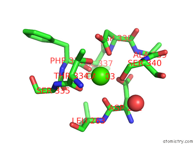
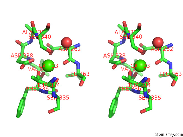
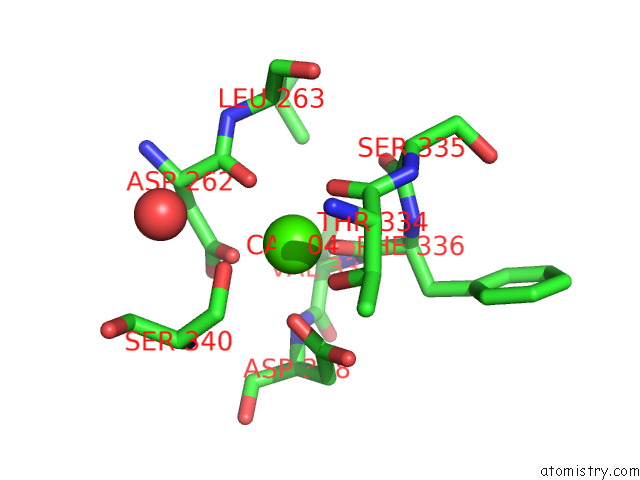
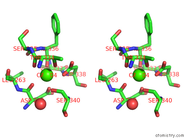
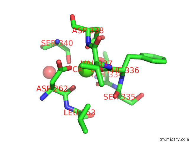
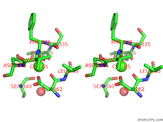
Calcium binding site 1 out of 8 in 7oih
Go back to
Calcium binding site 1 out
of 8 in the Glycosylation in the Crystal Structure of Neutrophil Myeloperoxidase
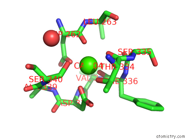
Mono view
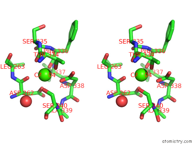
Stereo pair view
Mono view
Stereo pair view
A full contact list of Calcium with other atoms in the Ca binding
site number 1 of Glycosylation in the Crystal Structure of Neutrophil Myeloperoxidase within 5.0Å range:
|
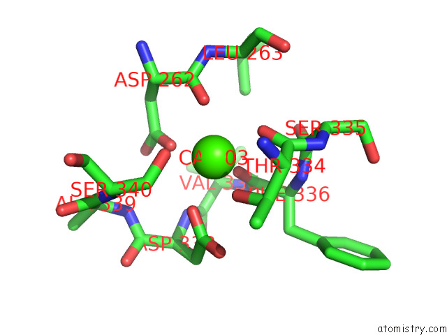
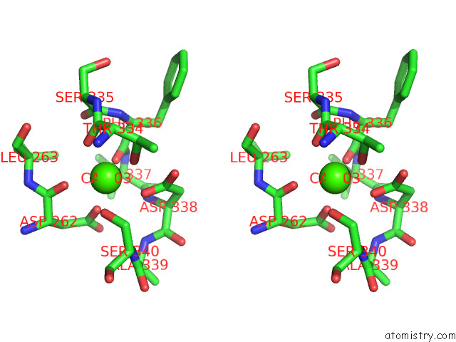
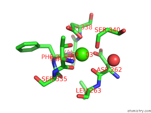
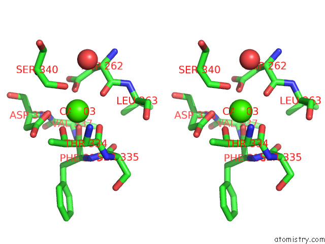
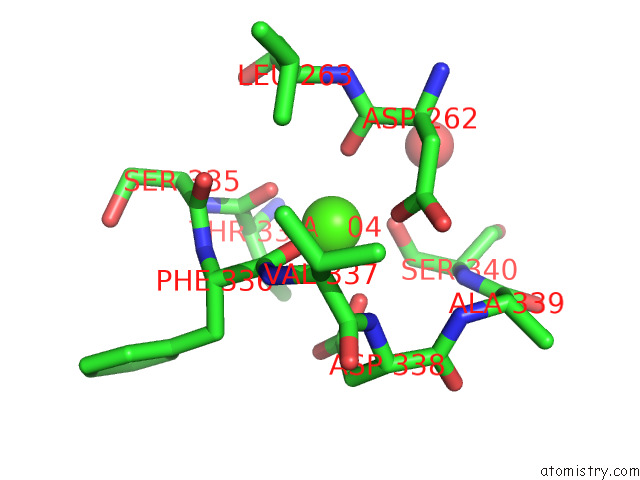
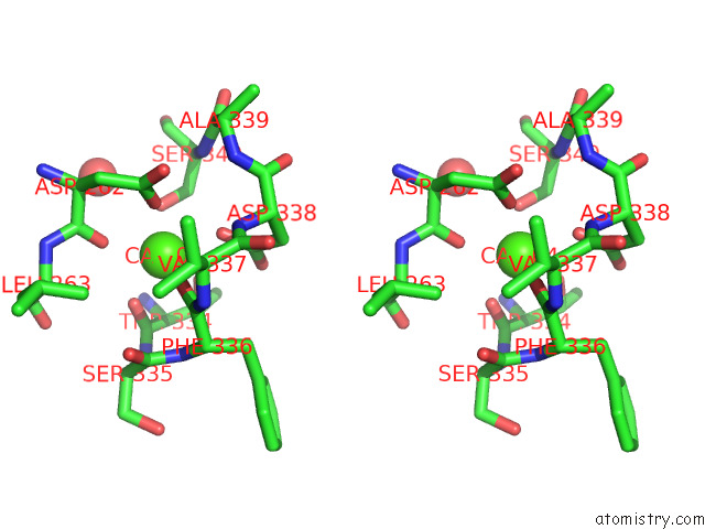
Calcium binding site 2 out of 8 in 7oih
Go back to
Calcium binding site 2 out
of 8 in the Glycosylation in the Crystal Structure of Neutrophil Myeloperoxidase
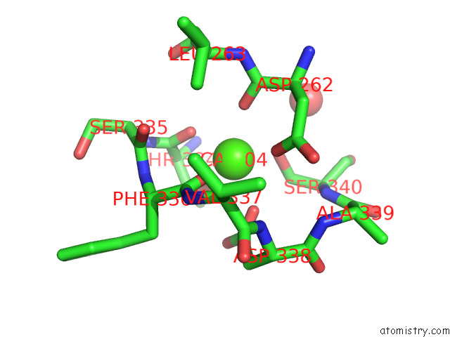
Mono view
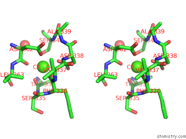
Stereo pair view
Mono view
Stereo pair view
A full contact list of Calcium with other atoms in the Ca binding
site number 2 of Glycosylation in the Crystal Structure of Neutrophil Myeloperoxidase within 5.0Å range:
|
Calcium binding site 3 out of 8 in 7oih
Go back to
Calcium binding site 3 out
of 8 in the Glycosylation in the Crystal Structure of Neutrophil Myeloperoxidase
Mono view
Stereo pair view
Mono view
Stereo pair view
A full contact list of Calcium with other atoms in the Ca binding
site number 3 of Glycosylation in the Crystal Structure of Neutrophil Myeloperoxidase within 5.0Å range:
|
Calcium binding site 4 out of 8 in 7oih
Go back to
Calcium binding site 4 out
of 8 in the Glycosylation in the Crystal Structure of Neutrophil Myeloperoxidase
Mono view
Stereo pair view
Mono view
Stereo pair view
A full contact list of Calcium with other atoms in the Ca binding
site number 4 of Glycosylation in the Crystal Structure of Neutrophil Myeloperoxidase within 5.0Å range:
|
Calcium binding site 5 out of 8 in 7oih
Go back to
Calcium binding site 5 out
of 8 in the Glycosylation in the Crystal Structure of Neutrophil Myeloperoxidase
Mono view
Stereo pair view
Mono view
Stereo pair view
A full contact list of Calcium with other atoms in the Ca binding
site number 5 of Glycosylation in the Crystal Structure of Neutrophil Myeloperoxidase within 5.0Å range:
|
Calcium binding site 6 out of 8 in 7oih
Go back to
Calcium binding site 6 out
of 8 in the Glycosylation in the Crystal Structure of Neutrophil Myeloperoxidase
Mono view
Stereo pair view
Mono view
Stereo pair view
A full contact list of Calcium with other atoms in the Ca binding
site number 6 of Glycosylation in the Crystal Structure of Neutrophil Myeloperoxidase within 5.0Å range:
|
Calcium binding site 7 out of 8 in 7oih
Go back to
Calcium binding site 7 out
of 8 in the Glycosylation in the Crystal Structure of Neutrophil Myeloperoxidase
Mono view
Stereo pair view
Mono view
Stereo pair view
A full contact list of Calcium with other atoms in the Ca binding
site number 7 of Glycosylation in the Crystal Structure of Neutrophil Myeloperoxidase within 5.0Å range:
|
Calcium binding site 8 out of 8 in 7oih
Go back to
Calcium binding site 8 out
of 8 in the Glycosylation in the Crystal Structure of Neutrophil Myeloperoxidase
Mono view
Stereo pair view
Mono view
Stereo pair view
A full contact list of Calcium with other atoms in the Ca binding
site number 8 of Glycosylation in the Crystal Structure of Neutrophil Myeloperoxidase within 5.0Å range:
|
Reference:
L.Krawczyk,
S.Semwal,
J.Soubhye,
S.Lemri Ouadriri,
M.Prevost,
P.Van Antwerpen,
G.Roos,
J.Bouckaert.
Native Glycosylation and Binding of the Antidepressant Paroxetine in A Low-Resolution Crystal Structure of Human Myeloperoxidase. Acta Crystallogr D Struct V. 78 1099 2022BIOL.
ISSN: ISSN 2059-7983
PubMed: 36048150
DOI: 10.1107/S2059798322007082
Page generated: Fri Jul 19 02:30:33 2024
ISSN: ISSN 2059-7983
PubMed: 36048150
DOI: 10.1107/S2059798322007082
Last articles
Zn in 9J0NZn in 9J0O
Zn in 9J0P
Zn in 9FJX
Zn in 9EKB
Zn in 9C0F
Zn in 9CAH
Zn in 9CH0
Zn in 9CH3
Zn in 9CH1