Calcium in PDB 7pug: GH115 Alpha-1,2-Glucuronidase in Complex with Xylopentaose
Enzymatic activity of GH115 Alpha-1,2-Glucuronidase in Complex with Xylopentaose
All present enzymatic activity of GH115 Alpha-1,2-Glucuronidase in Complex with Xylopentaose:
3.2.1.131;
3.2.1.131;
Protein crystallography data
The structure of GH115 Alpha-1,2-Glucuronidase in Complex with Xylopentaose, PDB code: 7pug
was solved by
C.Wilkens,
J.P.Morth,
I.Polikarpov,
with X-Ray Crystallography technique. A brief refinement statistics is given in the table below:
| Resolution Low / High (Å) | 65.26 / 2.66 |
| Space group | P 61 2 2 |
| Cell size a, b, c (Å), α, β, γ (°) | 148.63, 148.63, 272.79, 90, 90, 120 |
| R / Rfree (%) | 17.4 / 21 |
Other elements in 7pug:
The structure of GH115 Alpha-1,2-Glucuronidase in Complex with Xylopentaose also contains other interesting chemical elements:
| Chlorine | (Cl) | 13 atoms |
Calcium Binding Sites:
The binding sites of Calcium atom in the GH115 Alpha-1,2-Glucuronidase in Complex with Xylopentaose
(pdb code 7pug). This binding sites where shown within
5.0 Angstroms radius around Calcium atom.
In total 3 binding sites of Calcium where determined in the GH115 Alpha-1,2-Glucuronidase in Complex with Xylopentaose, PDB code: 7pug:
Jump to Calcium binding site number: 1; 2; 3;
In total 3 binding sites of Calcium where determined in the GH115 Alpha-1,2-Glucuronidase in Complex with Xylopentaose, PDB code: 7pug:
Jump to Calcium binding site number: 1; 2; 3;
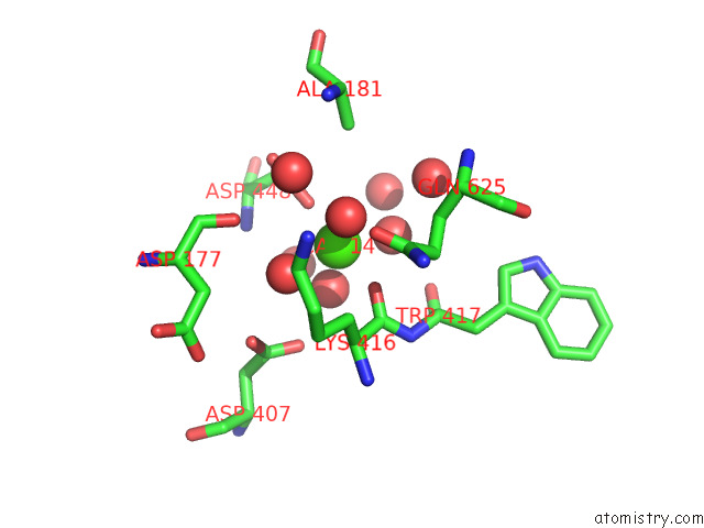
Calcium binding site 1 out of 3 in 7pug
Go back to
Calcium binding site 1 out
of 3 in the GH115 Alpha-1,2-Glucuronidase in Complex with Xylopentaose
Mono view

Stereo pair view
Mono view
Stereo pair view
A full contact list of Calcium with other atoms in the Ca binding
site number 1 of GH115 Alpha-1,2-Glucuronidase in Complex with Xylopentaose within 5.0Å range:
|
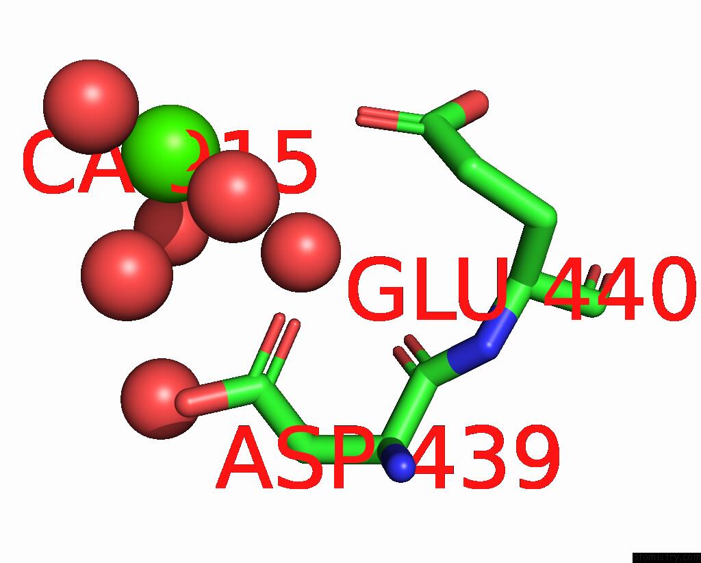
Calcium binding site 2 out of 3 in 7pug
Go back to
Calcium binding site 2 out
of 3 in the GH115 Alpha-1,2-Glucuronidase in Complex with Xylopentaose
Mono view
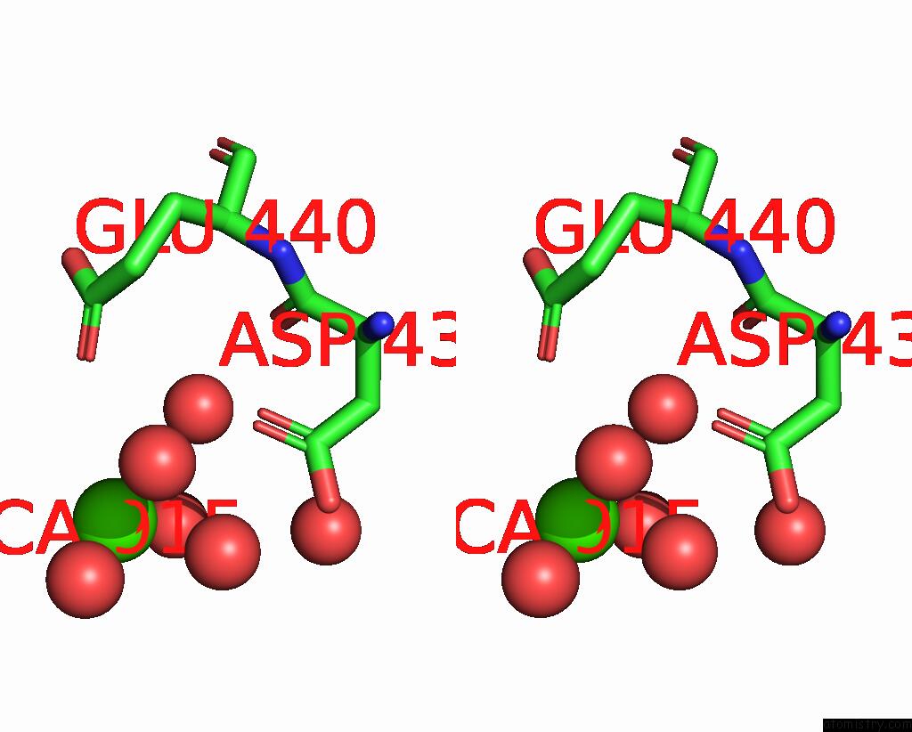
Stereo pair view
Mono view
Stereo pair view
A full contact list of Calcium with other atoms in the Ca binding
site number 2 of GH115 Alpha-1,2-Glucuronidase in Complex with Xylopentaose within 5.0Å range:
|
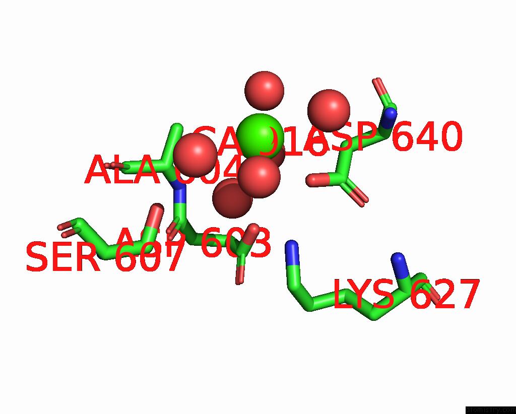
Calcium binding site 3 out of 3 in 7pug
Go back to
Calcium binding site 3 out
of 3 in the GH115 Alpha-1,2-Glucuronidase in Complex with Xylopentaose
Mono view

Stereo pair view
Mono view
Stereo pair view
A full contact list of Calcium with other atoms in the Ca binding
site number 3 of GH115 Alpha-1,2-Glucuronidase in Complex with Xylopentaose within 5.0Å range:
|
Reference:
C.Wilkens,
M.Vuillemin,
B.Pilgaard,
I.Polikarpov,
J.P.Morth.
A GH115 Alpha-Glucuronidase Structure Reveals Dimerization-Mediated Substrate Binding and A Proton Wire Potentially Important For Catalysis. Acta Crystallogr D Struct V. 78 658 2022BIOL.
ISSN: ISSN 2059-7983
PubMed: 35503213
DOI: 10.1107/S2059798322003527
Page generated: Fri Jul 19 03:18:33 2024
ISSN: ISSN 2059-7983
PubMed: 35503213
DOI: 10.1107/S2059798322003527
Last articles
Zn in 9J0NZn in 9J0O
Zn in 9J0P
Zn in 9FJX
Zn in 9EKB
Zn in 9C0F
Zn in 9CAH
Zn in 9CH0
Zn in 9CH3
Zn in 9CH1