Calcium in PDB 7q2a: Crystal Structure of Aphc in Complex with 4-Ethylcatechol
Enzymatic activity of Crystal Structure of Aphc in Complex with 4-Ethylcatechol
All present enzymatic activity of Crystal Structure of Aphc in Complex with 4-Ethylcatechol:
1.13.11.2;
1.13.11.2;
Protein crystallography data
The structure of Crystal Structure of Aphc in Complex with 4-Ethylcatechol, PDB code: 7q2a
was solved by
M.Zahn,
J.C.Grigg,
L.D.Eltis,
J.E.Mcgeehan,
with X-Ray Crystallography technique. A brief refinement statistics is given in the table below:
| Resolution Low / High (Å) | 47.19 / 1.60 |
| Space group | P 1 21 1 |
| Cell size a, b, c (Å), α, β, γ (°) | 81.58, 119.351, 84.528, 90, 114.56, 90 |
| R / Rfree (%) | 17.9 / 20.9 |
Other elements in 7q2a:
The structure of Crystal Structure of Aphc in Complex with 4-Ethylcatechol also contains other interesting chemical elements:
| Iron | (Fe) | 4 atoms |
Calcium Binding Sites:
The binding sites of Calcium atom in the Crystal Structure of Aphc in Complex with 4-Ethylcatechol
(pdb code 7q2a). This binding sites where shown within
5.0 Angstroms radius around Calcium atom.
In total 6 binding sites of Calcium where determined in the Crystal Structure of Aphc in Complex with 4-Ethylcatechol, PDB code: 7q2a:
Jump to Calcium binding site number: 1; 2; 3; 4; 5; 6;
In total 6 binding sites of Calcium where determined in the Crystal Structure of Aphc in Complex with 4-Ethylcatechol, PDB code: 7q2a:
Jump to Calcium binding site number: 1; 2; 3; 4; 5; 6;
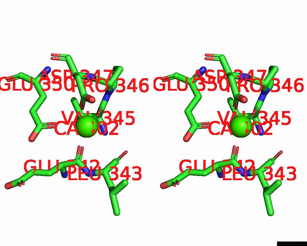
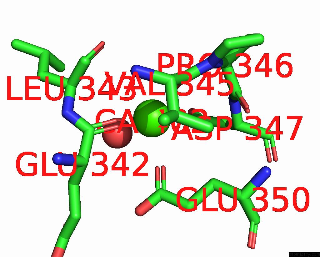
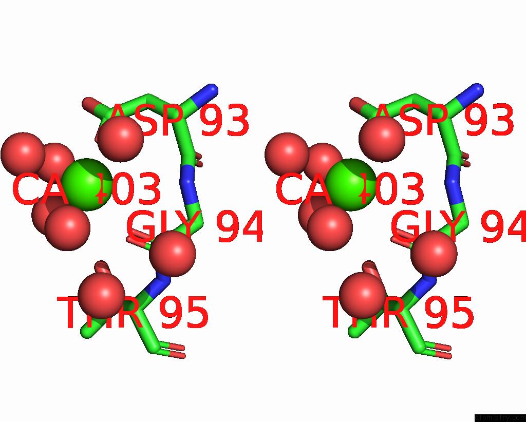
Calcium binding site 1 out of 6 in 7q2a
Go back to
Calcium binding site 1 out
of 6 in the Crystal Structure of Aphc in Complex with 4-Ethylcatechol

Mono view
Stereo pair view
Mono view
Stereo pair view
A full contact list of Calcium with other atoms in the Ca binding
site number 1 of Crystal Structure of Aphc in Complex with 4-Ethylcatechol within 5.0Å range:
|
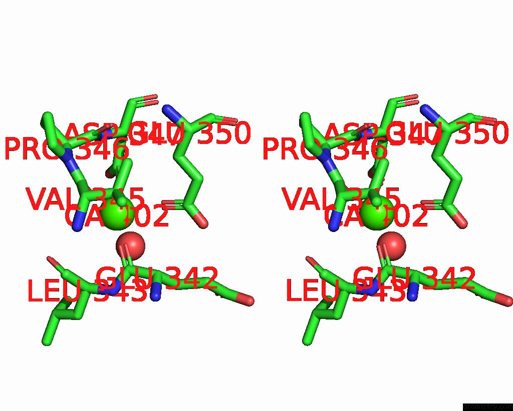
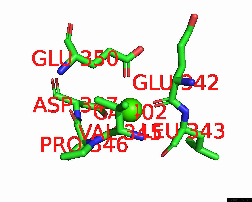
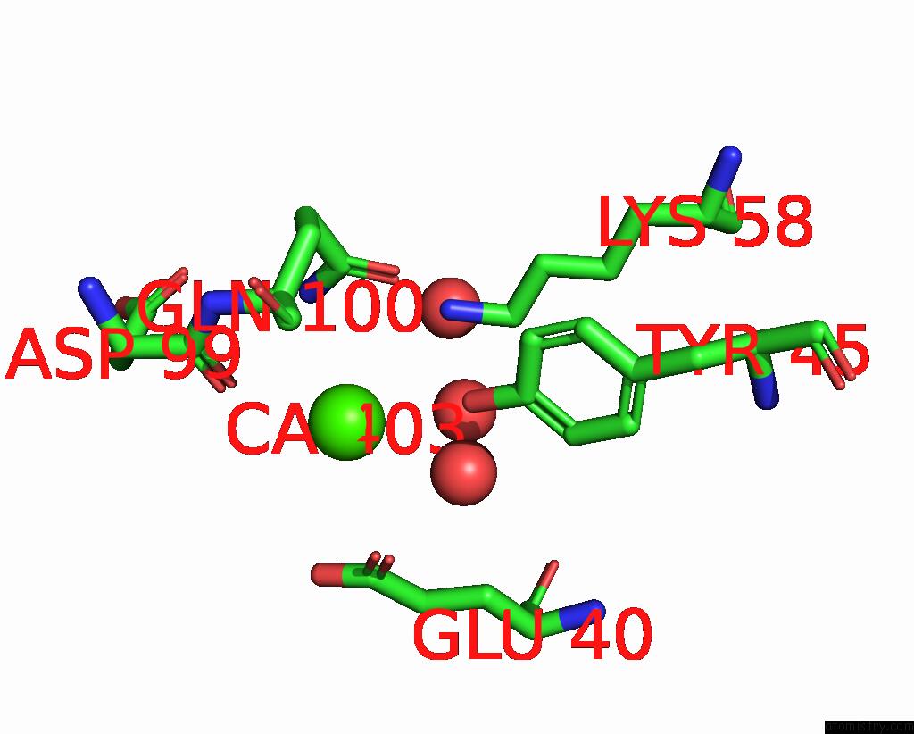
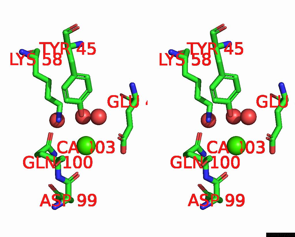
Calcium binding site 2 out of 6 in 7q2a
Go back to
Calcium binding site 2 out
of 6 in the Crystal Structure of Aphc in Complex with 4-Ethylcatechol
Mono view
Stereo pair view
Mono view
Stereo pair view
A full contact list of Calcium with other atoms in the Ca binding
site number 2 of Crystal Structure of Aphc in Complex with 4-Ethylcatechol within 5.0Å range:
|
Calcium binding site 3 out of 6 in 7q2a
Go back to
Calcium binding site 3 out
of 6 in the Crystal Structure of Aphc in Complex with 4-Ethylcatechol
Mono view

Stereo pair view
Mono view
Stereo pair view
A full contact list of Calcium with other atoms in the Ca binding
site number 3 of Crystal Structure of Aphc in Complex with 4-Ethylcatechol within 5.0Å range:
|
Calcium binding site 4 out of 6 in 7q2a
Go back to
Calcium binding site 4 out
of 6 in the Crystal Structure of Aphc in Complex with 4-Ethylcatechol

Mono view
Stereo pair view
Mono view
Stereo pair view
A full contact list of Calcium with other atoms in the Ca binding
site number 4 of Crystal Structure of Aphc in Complex with 4-Ethylcatechol within 5.0Å range:
|
Calcium binding site 5 out of 6 in 7q2a
Go back to
Calcium binding site 5 out
of 6 in the Crystal Structure of Aphc in Complex with 4-Ethylcatechol

Mono view

Stereo pair view
Mono view
Stereo pair view
A full contact list of Calcium with other atoms in the Ca binding
site number 5 of Crystal Structure of Aphc in Complex with 4-Ethylcatechol within 5.0Å range:
|
Calcium binding site 6 out of 6 in 7q2a
Go back to
Calcium binding site 6 out
of 6 in the Crystal Structure of Aphc in Complex with 4-Ethylcatechol
Mono view
Stereo pair view
Mono view
Stereo pair view
A full contact list of Calcium with other atoms in the Ca binding
site number 6 of Crystal Structure of Aphc in Complex with 4-Ethylcatechol within 5.0Å range:
|
Reference:
L.E.Navas,
M.Zahn,
H.Bajwa,
J.C.Grigg,
M.E.Wolf,
A.C.K.Chan,
M.E.P.Murphy,
J.E.Mcgeehan,
L.D.Eltis.
Characterization of A Phylogenetically Distinct Extradiol Dioxygenase Involved in the Bacterial Catabolism of Lignin-Derived Aromatic Compounds. J.Biol.Chem. V. 298 01871 2022.
ISSN: ESSN 1083-351X
PubMed: 35346686
DOI: 10.1016/J.JBC.2022.101871
Page generated: Fri Jul 19 03:23:20 2024
ISSN: ESSN 1083-351X
PubMed: 35346686
DOI: 10.1016/J.JBC.2022.101871
Last articles
Zn in 9J0NZn in 9J0O
Zn in 9J0P
Zn in 9FJX
Zn in 9EKB
Zn in 9C0F
Zn in 9CAH
Zn in 9CH0
Zn in 9CH3
Zn in 9CH1