Calcium in PDB 7s8d: Structure of Dna-Free Sgrai
Protein crystallography data
The structure of Structure of Dna-Free Sgrai, PDB code: 7s8d
was solved by
N.C.Horton,
with X-Ray Crystallography technique. A brief refinement statistics is given in the table below:
| Resolution Low / High (Å) | 38.00 / 2.02 |
| Space group | P 1 21 1 |
| Cell size a, b, c (Å), α, β, γ (°) | 70.11, 72.379, 78.64, 90, 107.95, 90 |
| R / Rfree (%) | 20.2 / 24.6 |
Calcium Binding Sites:
The binding sites of Calcium atom in the Structure of Dna-Free Sgrai
(pdb code 7s8d). This binding sites where shown within
5.0 Angstroms radius around Calcium atom.
In total 2 binding sites of Calcium where determined in the Structure of Dna-Free Sgrai, PDB code: 7s8d:
Jump to Calcium binding site number: 1; 2;
In total 2 binding sites of Calcium where determined in the Structure of Dna-Free Sgrai, PDB code: 7s8d:
Jump to Calcium binding site number: 1; 2;
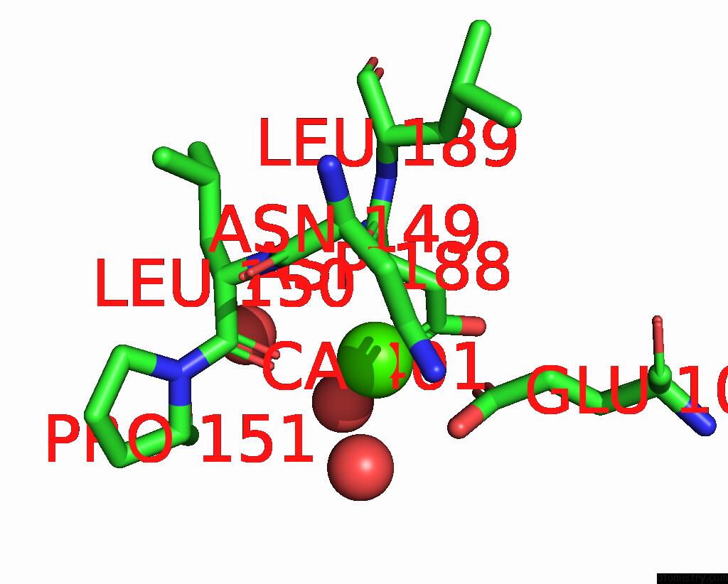
Calcium binding site 1 out of 2 in 7s8d
Go back to
Calcium binding site 1 out
of 2 in the Structure of Dna-Free Sgrai

Mono view
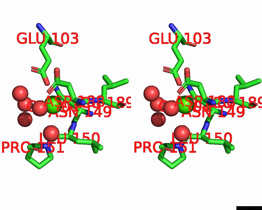
Stereo pair view
Mono view
Stereo pair view
A full contact list of Calcium with other atoms in the Ca binding
site number 1 of Structure of Dna-Free Sgrai within 5.0Å range:
|
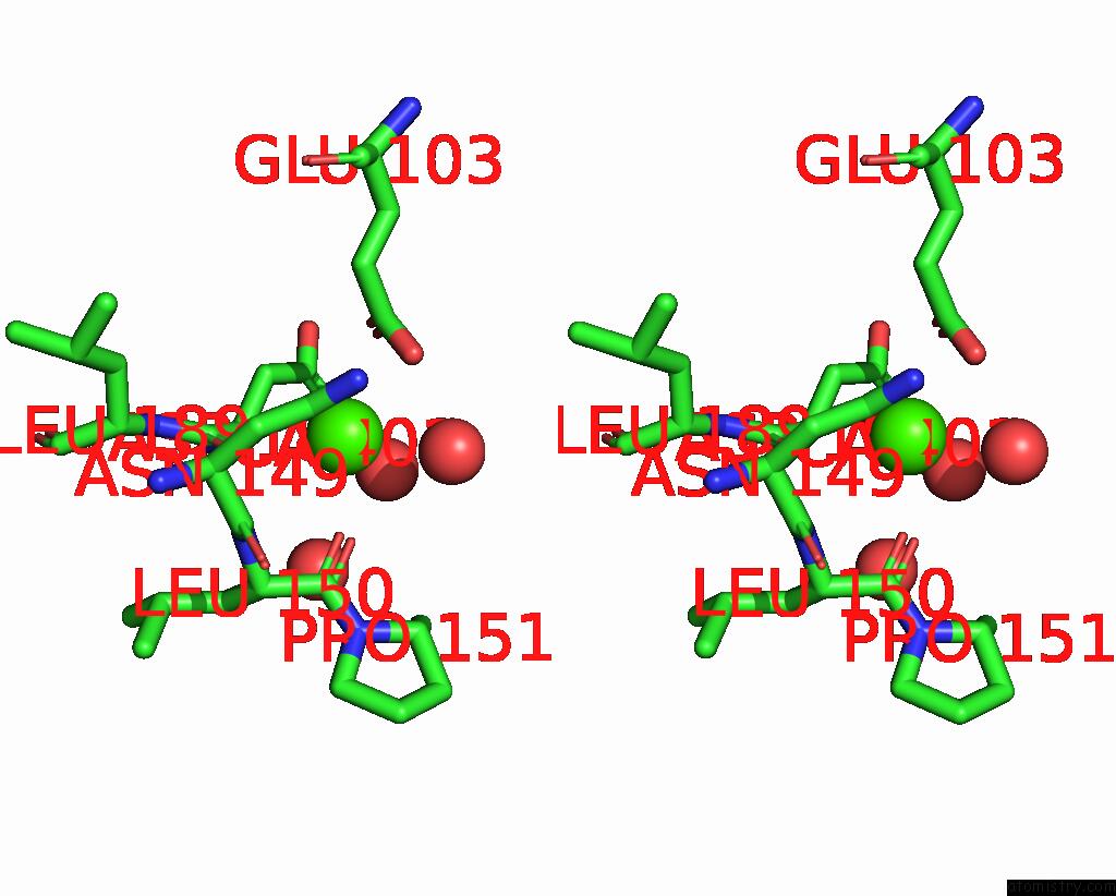
Calcium binding site 2 out of 2 in 7s8d
Go back to
Calcium binding site 2 out
of 2 in the Structure of Dna-Free Sgrai
Mono view
Stereo pair view
Mono view
Stereo pair view
A full contact list of Calcium with other atoms in the Ca binding
site number 2 of Structure of Dna-Free Sgrai within 5.0Å range:
|
Reference:
Z.Shan,
N.Ghadirian,
D.Lyumkis,
N.C.Horton.
Pretransition State and Apo Structures of the Filament-Forming Enzyme Sgrai Elucidate Mechanisms of Activation and Substrate Specificity. J.Biol.Chem. V. 298 01760 2022.
ISSN: ESSN 1083-351X
PubMed: 35202658
DOI: 10.1016/J.JBC.2022.101760
Page generated: Fri Jul 19 03:58:56 2024
ISSN: ESSN 1083-351X
PubMed: 35202658
DOI: 10.1016/J.JBC.2022.101760
Last articles
Zn in 9J0NZn in 9J0O
Zn in 9J0P
Zn in 9FJX
Zn in 9EKB
Zn in 9C0F
Zn in 9CAH
Zn in 9CH0
Zn in 9CH3
Zn in 9CH1