Calcium in PDB 7sc3: Crystal Structure of the N-Domain of Cardiac Muscle Troponin C Tethered to the Switch Region of Cardiac Muscle Troponin I (Orthorhombic Form)
Protein crystallography data
The structure of Crystal Structure of the N-Domain of Cardiac Muscle Troponin C Tethered to the Switch Region of Cardiac Muscle Troponin I (Orthorhombic Form), PDB code: 7sc3
was solved by
J.S.Sack,
with X-Ray Crystallography technique. A brief refinement statistics is given in the table below:
| Resolution Low / High (Å) | 34.95 / 2.23 |
| Space group | P 21 21 21 |
| Cell size a, b, c (Å), α, β, γ (°) | 38.849, 40.958, 66.997, 90, 90, 90 |
| R / Rfree (%) | 24.3 / 27.8 |
Calcium Binding Sites:
The binding sites of Calcium atom in the Crystal Structure of the N-Domain of Cardiac Muscle Troponin C Tethered to the Switch Region of Cardiac Muscle Troponin I (Orthorhombic Form)
(pdb code 7sc3). This binding sites where shown within
5.0 Angstroms radius around Calcium atom.
In total only one binding site of Calcium was determined in the Crystal Structure of the N-Domain of Cardiac Muscle Troponin C Tethered to the Switch Region of Cardiac Muscle Troponin I (Orthorhombic Form), PDB code: 7sc3:
In total only one binding site of Calcium was determined in the Crystal Structure of the N-Domain of Cardiac Muscle Troponin C Tethered to the Switch Region of Cardiac Muscle Troponin I (Orthorhombic Form), PDB code: 7sc3:
Calcium binding site 1 out of 1 in 7sc3
Go back to
Calcium binding site 1 out
of 1 in the Crystal Structure of the N-Domain of Cardiac Muscle Troponin C Tethered to the Switch Region of Cardiac Muscle Troponin I (Orthorhombic Form)
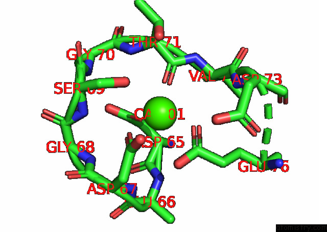
Mono view
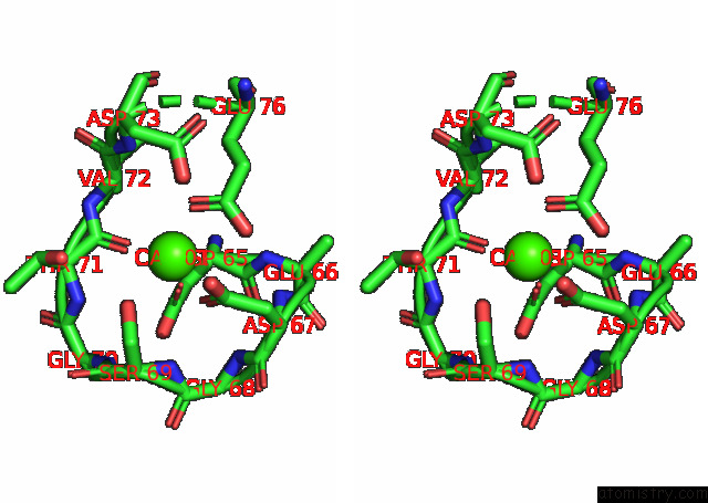
Stereo pair view
Mono view
Stereo pair view
A full contact list of Calcium with other atoms in the Ca binding
site number 1 of Crystal Structure of the N-Domain of Cardiac Muscle Troponin C Tethered to the Switch Region of Cardiac Muscle Troponin I (Orthorhombic Form) within 5.0Å range:
|
Reference:
C.Yan,
J.S.Sack.
X-Ray Structure of A Human Cardiac Muscle Troponin C / Troponin I Chimera in Two Crystal Forms Acta Crystallogr.,Sect.F 2021.
ISSN: ESSN 2053-230X
Page generated: Fri Jul 19 03:59:44 2024
ISSN: ESSN 2053-230X
Last articles
Zn in 9J0NZn in 9J0O
Zn in 9J0P
Zn in 9FJX
Zn in 9EKB
Zn in 9C0F
Zn in 9CAH
Zn in 9CH0
Zn in 9CH3
Zn in 9CH1