Calcium in PDB 7vo7: Crystal Structure of Trypsin in Complex with Lima Bean Trypsin Inhibitor at 2.25A Resolution.
Enzymatic activity of Crystal Structure of Trypsin in Complex with Lima Bean Trypsin Inhibitor at 2.25A Resolution.
All present enzymatic activity of Crystal Structure of Trypsin in Complex with Lima Bean Trypsin Inhibitor at 2.25A Resolution.:
3.4.21.4;
3.4.21.4;
Protein crystallography data
The structure of Crystal Structure of Trypsin in Complex with Lima Bean Trypsin Inhibitor at 2.25A Resolution., PDB code: 7vo7
was solved by
M.S.Ahmad,
Z.Akbar,
M.I.Choudhary,
with X-Ray Crystallography technique. A brief refinement statistics is given in the table below:
| Resolution Low / High (Å) | 17.91 / 2.25 |
| Space group | P 41 |
| Cell size a, b, c (Å), α, β, γ (°) | 54.009, 54.009, 179.817, 90, 90, 90 |
| R / Rfree (%) | 18.1 / 23.3 |
Other elements in 7vo7:
The structure of Crystal Structure of Trypsin in Complex with Lima Bean Trypsin Inhibitor at 2.25A Resolution. also contains other interesting chemical elements:
| Chlorine | (Cl) | 2 atoms |
| Sodium | (Na) | 1 atom |
Calcium Binding Sites:
The binding sites of Calcium atom in the Crystal Structure of Trypsin in Complex with Lima Bean Trypsin Inhibitor at 2.25A Resolution.
(pdb code 7vo7). This binding sites where shown within
5.0 Angstroms radius around Calcium atom.
In total 2 binding sites of Calcium where determined in the Crystal Structure of Trypsin in Complex with Lima Bean Trypsin Inhibitor at 2.25A Resolution., PDB code: 7vo7:
Jump to Calcium binding site number: 1; 2;
In total 2 binding sites of Calcium where determined in the Crystal Structure of Trypsin in Complex with Lima Bean Trypsin Inhibitor at 2.25A Resolution., PDB code: 7vo7:
Jump to Calcium binding site number: 1; 2;
Calcium binding site 1 out of 2 in 7vo7
Go back to
Calcium binding site 1 out
of 2 in the Crystal Structure of Trypsin in Complex with Lima Bean Trypsin Inhibitor at 2.25A Resolution.
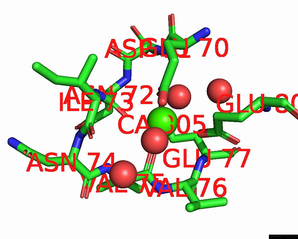
Mono view
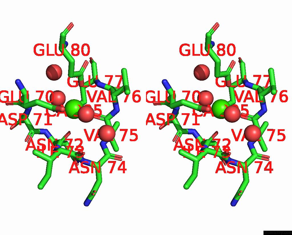
Stereo pair view
Mono view
Stereo pair view
A full contact list of Calcium with other atoms in the Ca binding
site number 1 of Crystal Structure of Trypsin in Complex with Lima Bean Trypsin Inhibitor at 2.25A Resolution. within 5.0Å range:
|
Calcium binding site 2 out of 2 in 7vo7
Go back to
Calcium binding site 2 out
of 2 in the Crystal Structure of Trypsin in Complex with Lima Bean Trypsin Inhibitor at 2.25A Resolution.
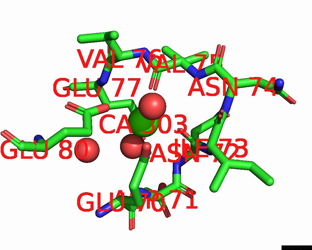
Mono view
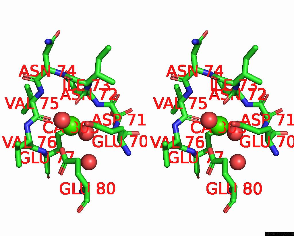
Stereo pair view
Mono view
Stereo pair view
A full contact list of Calcium with other atoms in the Ca binding
site number 2 of Crystal Structure of Trypsin in Complex with Lima Bean Trypsin Inhibitor at 2.25A Resolution. within 5.0Å range:
|
Reference:
M.S.Ahmad,
Z.Akbar,
M.I.Choudhary.
Insight Into the Structural Basis of the Dual Inhibitory Mode of Lima Bean (Phaseolus Lunatus) Serine Protease Inhibitor. Proteins V. 91 22 2023.
ISSN: ESSN 1097-0134
PubMed: 35927030
DOI: 10.1002/PROT.26407
Page generated: Fri Jul 19 05:23:57 2024
ISSN: ESSN 1097-0134
PubMed: 35927030
DOI: 10.1002/PROT.26407
Last articles
Zn in 9MJ5Zn in 9HNW
Zn in 9G0L
Zn in 9FNE
Zn in 9DZN
Zn in 9E0I
Zn in 9D32
Zn in 9DAK
Zn in 8ZXC
Zn in 8ZUF