Calcium in PDB 7w4z: Crystal Structure of Fragmin Domain-1 in Complex with Actin (Amppnp- Form)
Protein crystallography data
The structure of Crystal Structure of Fragmin Domain-1 in Complex with Actin (Amppnp- Form), PDB code: 7w4z
was solved by
S.Takeda,
with X-Ray Crystallography technique. A brief refinement statistics is given in the table below:
| Resolution Low / High (Å) | 29.35 / 1.15 |
| Space group | P 21 21 21 |
| Cell size a, b, c (Å), α, β, γ (°) | 57.09, 91.07, 114.85, 90, 90, 90 |
| R / Rfree (%) | 14.3 / 15.8 |
Other elements in 7w4z:
The structure of Crystal Structure of Fragmin Domain-1 in Complex with Actin (Amppnp- Form) also contains other interesting chemical elements:
| Magnesium | (Mg) | 1 atom |
Calcium Binding Sites:
The binding sites of Calcium atom in the Crystal Structure of Fragmin Domain-1 in Complex with Actin (Amppnp- Form)
(pdb code 7w4z). This binding sites where shown within
5.0 Angstroms radius around Calcium atom.
In total 2 binding sites of Calcium where determined in the Crystal Structure of Fragmin Domain-1 in Complex with Actin (Amppnp- Form), PDB code: 7w4z:
Jump to Calcium binding site number: 1; 2;
In total 2 binding sites of Calcium where determined in the Crystal Structure of Fragmin Domain-1 in Complex with Actin (Amppnp- Form), PDB code: 7w4z:
Jump to Calcium binding site number: 1; 2;
Calcium binding site 1 out of 2 in 7w4z
Go back to
Calcium binding site 1 out
of 2 in the Crystal Structure of Fragmin Domain-1 in Complex with Actin (Amppnp- Form)
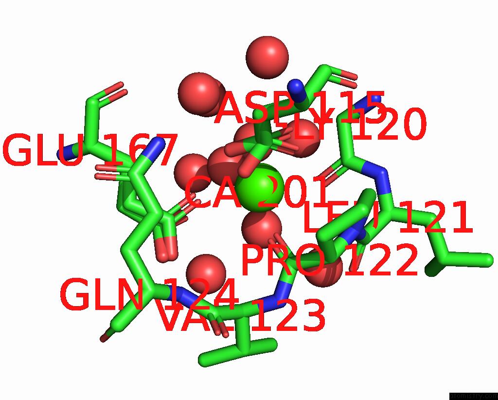
Mono view

Stereo pair view
Mono view
Stereo pair view
A full contact list of Calcium with other atoms in the Ca binding
site number 1 of Crystal Structure of Fragmin Domain-1 in Complex with Actin (Amppnp- Form) within 5.0Å range:
|
Calcium binding site 2 out of 2 in 7w4z
Go back to
Calcium binding site 2 out
of 2 in the Crystal Structure of Fragmin Domain-1 in Complex with Actin (Amppnp- Form)

Mono view
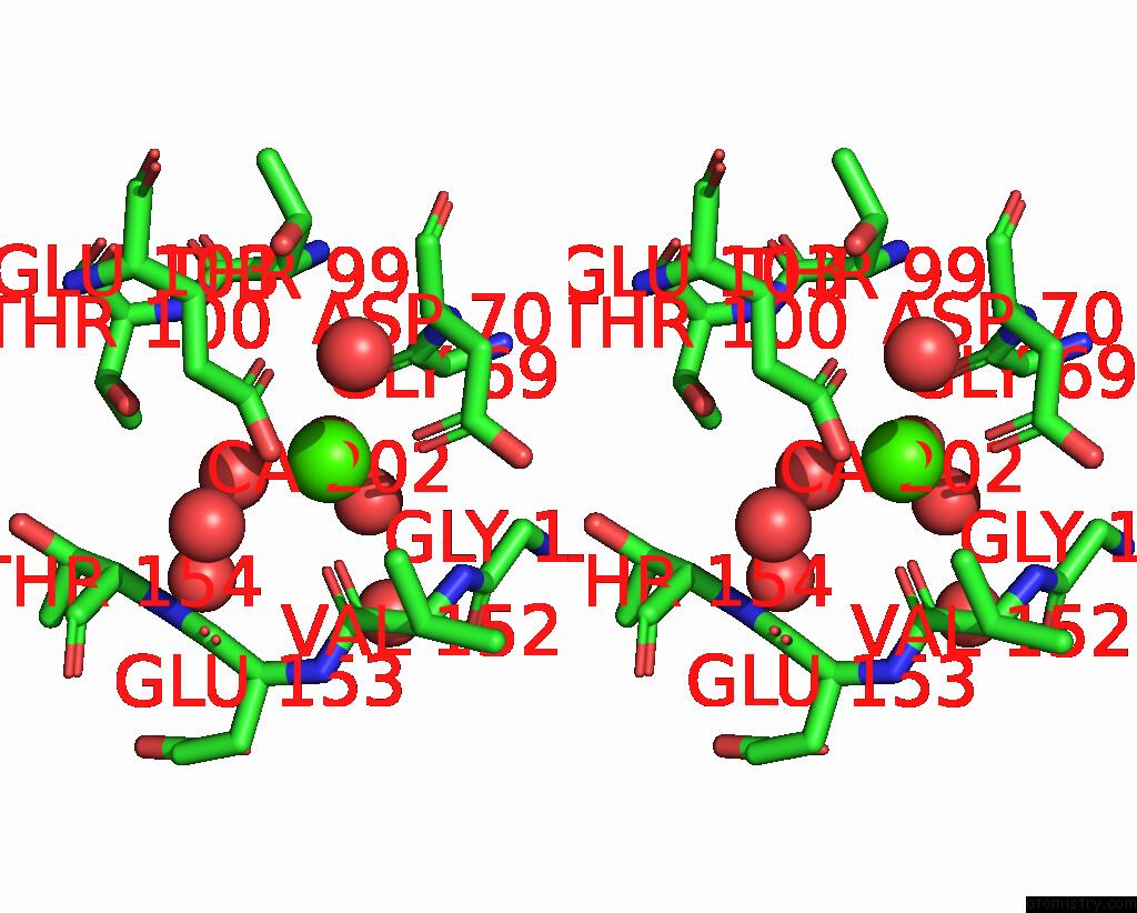
Stereo pair view
Mono view
Stereo pair view
A full contact list of Calcium with other atoms in the Ca binding
site number 2 of Crystal Structure of Fragmin Domain-1 in Complex with Actin (Amppnp- Form) within 5.0Å range:
|
Reference:
Y.Kanematsu,
A.Narita,
T.Oda,
R.Koike,
M.Ota,
Y.Takano,
K.Moritsugu,
I.Fujiwara,
K.Tanaka,
H.Komatsu,
T.Nagae,
N.Watanabe,
M.Iwasa,
Y.Maeda,
S.Takeda.
Structures and Mechanisms of Actin Atp Hydrolysis. Proc.Natl.Acad.Sci.Usa V. 119 41119 2022.
ISSN: ESSN 1091-6490
PubMed: 36252034
DOI: 10.1073/PNAS.2122641119
Page generated: Fri Jul 19 05:35:55 2024
ISSN: ESSN 1091-6490
PubMed: 36252034
DOI: 10.1073/PNAS.2122641119
Last articles
Zn in 9MJ5Zn in 9HNW
Zn in 9G0L
Zn in 9FNE
Zn in 9DZN
Zn in 9E0I
Zn in 9D32
Zn in 9DAK
Zn in 8ZXC
Zn in 8ZUF