Calcium in PDB 7y9o: Crystal Structure of A CYP109B4 Variant From Bacillus Sonorensis
Protein crystallography data
The structure of Crystal Structure of A CYP109B4 Variant From Bacillus Sonorensis, PDB code: 7y9o
was solved by
P.P.Shen,
J.-W.Huang,
X.Li,
W.D.Liu,
C.-C.Chen,
R.-T.Guo,
with X-Ray Crystallography technique. A brief refinement statistics is given in the table below:
| Resolution Low / High (Å) | 38.06 / 1.84 |
| Space group | P 21 21 21 |
| Cell size a, b, c (Å), α, β, γ (°) | 47.87, 62.602, 140.526, 90, 90, 90 |
| R / Rfree (%) | 19.1 / 22.2 |
Other elements in 7y9o:
The structure of Crystal Structure of A CYP109B4 Variant From Bacillus Sonorensis also contains other interesting chemical elements:
| Iron | (Fe) | 1 atom |
Calcium Binding Sites:
The binding sites of Calcium atom in the Crystal Structure of A CYP109B4 Variant From Bacillus Sonorensis
(pdb code 7y9o). This binding sites where shown within
5.0 Angstroms radius around Calcium atom.
In total 2 binding sites of Calcium where determined in the Crystal Structure of A CYP109B4 Variant From Bacillus Sonorensis, PDB code: 7y9o:
Jump to Calcium binding site number: 1; 2;
In total 2 binding sites of Calcium where determined in the Crystal Structure of A CYP109B4 Variant From Bacillus Sonorensis, PDB code: 7y9o:
Jump to Calcium binding site number: 1; 2;
Calcium binding site 1 out of 2 in 7y9o
Go back to
Calcium binding site 1 out
of 2 in the Crystal Structure of A CYP109B4 Variant From Bacillus Sonorensis

Mono view
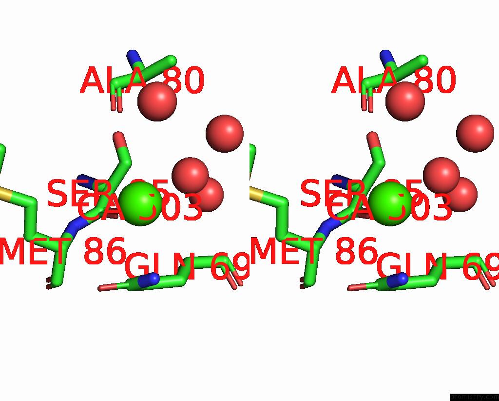
Stereo pair view
Mono view
Stereo pair view
A full contact list of Calcium with other atoms in the Ca binding
site number 1 of Crystal Structure of A CYP109B4 Variant From Bacillus Sonorensis within 5.0Å range:
|
Calcium binding site 2 out of 2 in 7y9o
Go back to
Calcium binding site 2 out
of 2 in the Crystal Structure of A CYP109B4 Variant From Bacillus Sonorensis
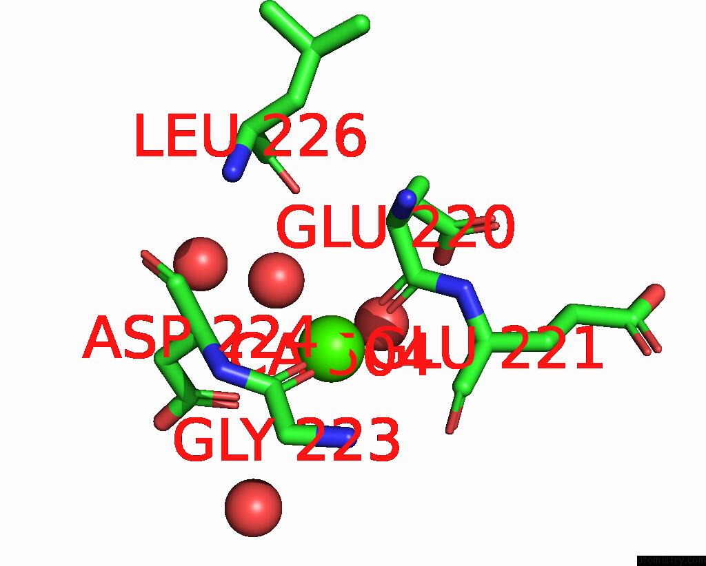
Mono view
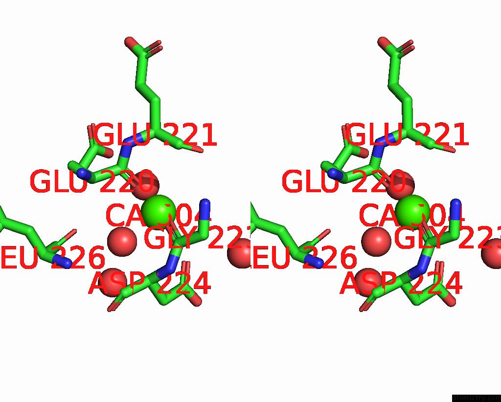
Stereo pair view
Mono view
Stereo pair view
A full contact list of Calcium with other atoms in the Ca binding
site number 2 of Crystal Structure of A CYP109B4 Variant From Bacillus Sonorensis within 5.0Å range:
|
Reference:
X.Zhang,
P.Shen,
J.Zhao,
Y.Chen,
X.Li,
J.W.Huang,
L.Zhang,
Q.Li,
C.Gao,
Q.Xing,
C.C.Chen,
R.T.Guo,
A.Li.
Rationally Controlling Selective Steroid Hydroxylation Via Scaffold Sampling of A P450 Family Acs Catalysis V. 13 1280 2023.
ISSN: ESSN 2155-5435
DOI: 10.1021/ACSCATAL.2C04906
Page generated: Fri Jul 19 06:12:10 2024
ISSN: ESSN 2155-5435
DOI: 10.1021/ACSCATAL.2C04906
Last articles
Zn in 9J0NZn in 9J0O
Zn in 9J0P
Zn in 9FJX
Zn in 9EKB
Zn in 9C0F
Zn in 9CAH
Zn in 9CH0
Zn in 9CH3
Zn in 9CH1