Calcium in PDB 7yyv: Molecular Snapshots of Drug Release From Tubulin: 1 Nanosecond After Photoactivation.
Protein crystallography data
The structure of Molecular Snapshots of Drug Release From Tubulin: 1 Nanosecond After Photoactivation., PDB code: 7yyv
was solved by
M.Wranik,
T.Weinert,
J.Standfuss,
with X-Ray Crystallography technique. A brief refinement statistics is given in the table below:
| Resolution Low / High (Å) | 9.49 / 2.20 |
| Space group | P 1 21 1 |
| Cell size a, b, c (Å), α, β, γ (°) | 74.53, 92.58, 83.99, 90, 96.71, 90 |
| R / Rfree (%) | 31.1 / 35.2 |
Other elements in 7yyv:
The structure of Molecular Snapshots of Drug Release From Tubulin: 1 Nanosecond After Photoactivation. also contains other interesting chemical elements:
| Magnesium | (Mg) | 1 atom |
Calcium Binding Sites:
The binding sites of Calcium atom in the Molecular Snapshots of Drug Release From Tubulin: 1 Nanosecond After Photoactivation.
(pdb code 7yyv). This binding sites where shown within
5.0 Angstroms radius around Calcium atom.
In total only one binding site of Calcium was determined in the Molecular Snapshots of Drug Release From Tubulin: 1 Nanosecond After Photoactivation., PDB code: 7yyv:
In total only one binding site of Calcium was determined in the Molecular Snapshots of Drug Release From Tubulin: 1 Nanosecond After Photoactivation., PDB code: 7yyv:
Calcium binding site 1 out of 1 in 7yyv
Go back to
Calcium binding site 1 out
of 1 in the Molecular Snapshots of Drug Release From Tubulin: 1 Nanosecond After Photoactivation.
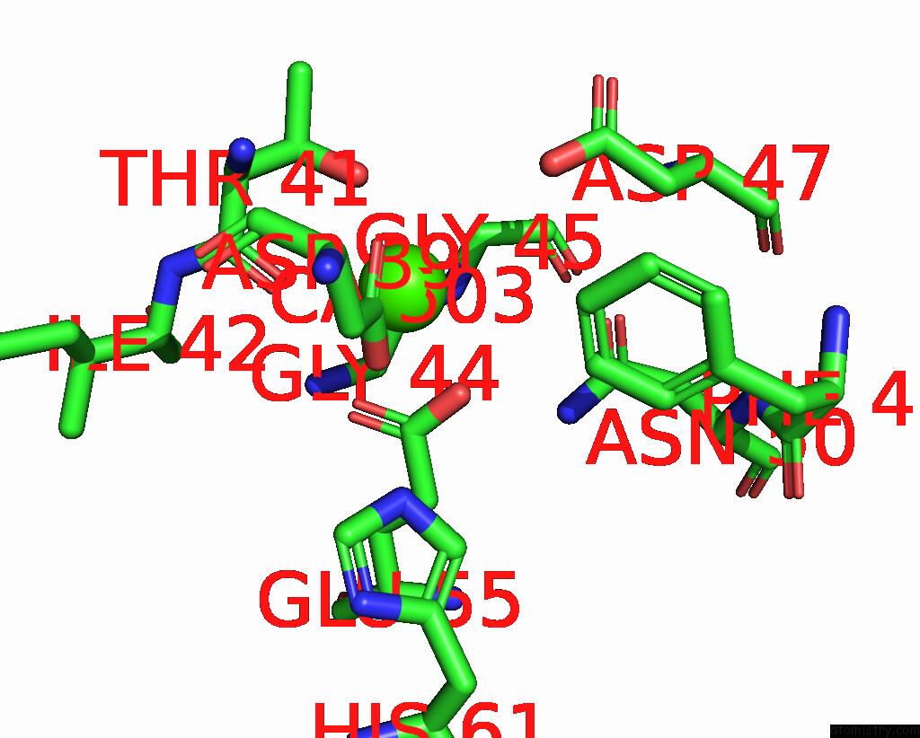
Mono view
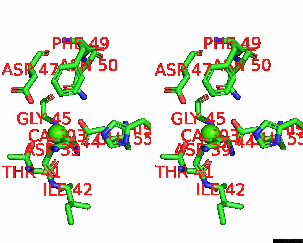
Stereo pair view
Mono view
Stereo pair view
A full contact list of Calcium with other atoms in the Ca binding
site number 1 of Molecular Snapshots of Drug Release From Tubulin: 1 Nanosecond After Photoactivation. within 5.0Å range:
|
Reference:
M.Wranik,
T.Weinert,
C.Slavov,
T.Masini,
A.Furrer,
N.Gaillard,
D.Gioia,
M.Ferrarotti,
D.James,
H.Glover,
M.Carrillo,
D.Kekilli,
R.Stipp,
P.Skopintsev,
S.Brunle,
T.Muhlethaler,
J.Beale,
D.Gashi,
K.Nass,
D.Ozerov,
P.Johnson,
C.Cirelli,
C.Bacellar,
M.Braun,
M.Wang,
F.Dworkowski,
C.Milne,
A.Cavalli,
J.Wachtveitl,
M.Steinmetz,
J.Standfuss.
Watching the Release of A Photopharmacological Drug From Tubulin Using Time-Resolved Serial Crystallography Nat Commun 2023.
ISSN: ESSN 2041-1723
DOI: 10.1038/S41467-023-36481-5
Page generated: Fri Jul 19 06:23:53 2024
ISSN: ESSN 2041-1723
DOI: 10.1038/S41467-023-36481-5
Last articles
Zn in 9JYWZn in 9IR4
Zn in 9IR3
Zn in 9GMX
Zn in 9GMW
Zn in 9JEJ
Zn in 9ERF
Zn in 9ERE
Zn in 9EGV
Zn in 9EGW