Calcium in PDB 8anv: Crystal Structure of PHI3T_93 and PHI3T Aimx Complex
Protein crystallography data
The structure of Crystal Structure of PHI3T_93 and PHI3T Aimx Complex, PDB code: 8anv
was solved by
S.Zamora-Caballero,
A.Marina,
with X-Ray Crystallography technique. A brief refinement statistics is given in the table below:
| Resolution Low / High (Å) | 47.67 / 2.20 |
| Space group | P 32 2 1 |
| Cell size a, b, c (Å), α, β, γ (°) | 66.438, 66.438, 85.129, 90, 90, 120 |
| R / Rfree (%) | 22.7 / 23.4 |
Other elements in 8anv:
The structure of Crystal Structure of PHI3T_93 and PHI3T Aimx Complex also contains other interesting chemical elements:
| Nickel | (Ni) | 1 atom |
Calcium Binding Sites:
The binding sites of Calcium atom in the Crystal Structure of PHI3T_93 and PHI3T Aimx Complex
(pdb code 8anv). This binding sites where shown within
5.0 Angstroms radius around Calcium atom.
In total only one binding site of Calcium was determined in the Crystal Structure of PHI3T_93 and PHI3T Aimx Complex, PDB code: 8anv:
In total only one binding site of Calcium was determined in the Crystal Structure of PHI3T_93 and PHI3T Aimx Complex, PDB code: 8anv:
Calcium binding site 1 out of 1 in 8anv
Go back to
Calcium binding site 1 out
of 1 in the Crystal Structure of PHI3T_93 and PHI3T Aimx Complex
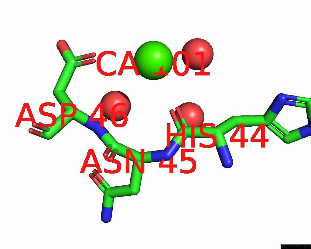
Mono view
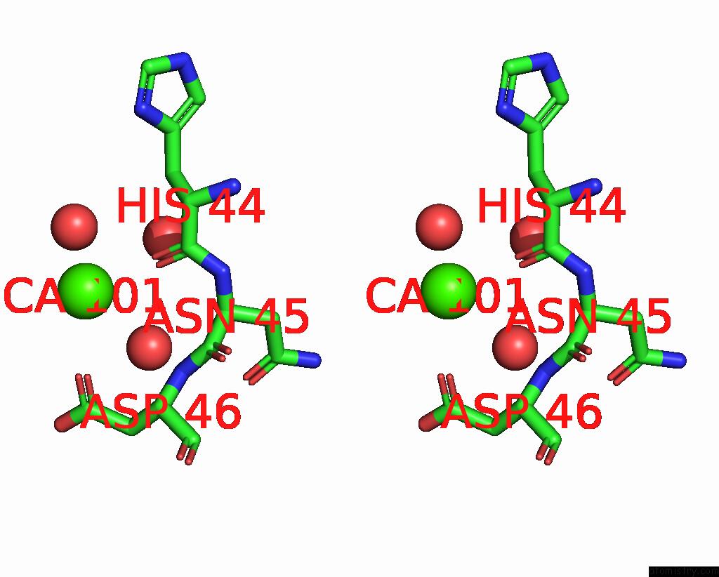
Stereo pair view
Mono view
Stereo pair view
A full contact list of Calcium with other atoms in the Ca binding
site number 1 of Crystal Structure of PHI3T_93 and PHI3T Aimx Complex within 5.0Å range:
|
Reference:
S.Zamora-Caballero,
A.Marina.
Antagonistic Interactions Between Phage and Host Factors Control Arbitrium Lysis-Lysogeny Decision Nat Microbiol 2023.
ISSN: ESSN 2058-5276
Page generated: Fri Jul 19 06:59:36 2024
ISSN: ESSN 2058-5276
Last articles
Zn in 9J0NZn in 9J0O
Zn in 9J0P
Zn in 9FJX
Zn in 9EKB
Zn in 9C0F
Zn in 9CAH
Zn in 9CH0
Zn in 9CH3
Zn in 9CH1