Calcium in PDB 8ax0: Crystal Structure of Trametes Versicolor Glutathione Transferase Omega 3S in Complex with Sodium Nitroprusside
Enzymatic activity of Crystal Structure of Trametes Versicolor Glutathione Transferase Omega 3S in Complex with Sodium Nitroprusside
All present enzymatic activity of Crystal Structure of Trametes Versicolor Glutathione Transferase Omega 3S in Complex with Sodium Nitroprusside:
2.5.1.18;
2.5.1.18;
Protein crystallography data
The structure of Crystal Structure of Trametes Versicolor Glutathione Transferase Omega 3S in Complex with Sodium Nitroprusside, PDB code: 8ax0
was solved by
M.Schwartz,
C.Didierjean,
with X-Ray Crystallography technique. A brief refinement statistics is given in the table below:
| Resolution Low / High (Å) | 47.74 / 1.65 |
| Space group | P 21 21 21 |
| Cell size a, b, c (Å), α, β, γ (°) | 50.51, 104.31, 107.38, 90, 90, 90 |
| R / Rfree (%) | 16.5 / 20.2 |
Other elements in 8ax0:
The structure of Crystal Structure of Trametes Versicolor Glutathione Transferase Omega 3S in Complex with Sodium Nitroprusside also contains other interesting chemical elements:
| Iron | (Fe) | 4 atoms |
Calcium Binding Sites:
The binding sites of Calcium atom in the Crystal Structure of Trametes Versicolor Glutathione Transferase Omega 3S in Complex with Sodium Nitroprusside
(pdb code 8ax0). This binding sites where shown within
5.0 Angstroms radius around Calcium atom.
In total only one binding site of Calcium was determined in the Crystal Structure of Trametes Versicolor Glutathione Transferase Omega 3S in Complex with Sodium Nitroprusside, PDB code: 8ax0:
In total only one binding site of Calcium was determined in the Crystal Structure of Trametes Versicolor Glutathione Transferase Omega 3S in Complex with Sodium Nitroprusside, PDB code: 8ax0:
Calcium binding site 1 out of 1 in 8ax0
Go back to
Calcium binding site 1 out
of 1 in the Crystal Structure of Trametes Versicolor Glutathione Transferase Omega 3S in Complex with Sodium Nitroprusside
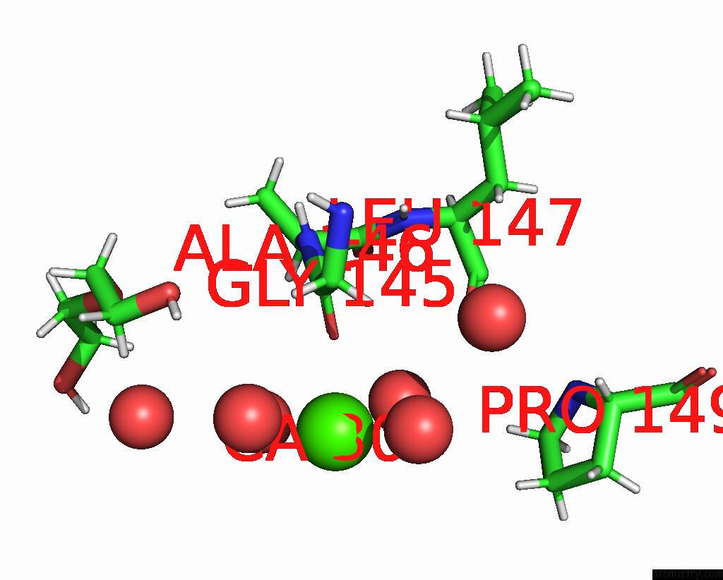
Mono view
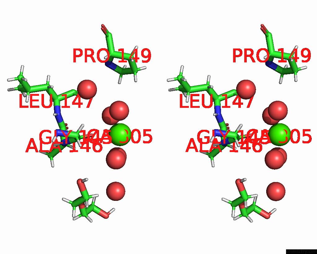
Stereo pair view
Mono view
Stereo pair view
A full contact list of Calcium with other atoms in the Ca binding
site number 1 of Crystal Structure of Trametes Versicolor Glutathione Transferase Omega 3S in Complex with Sodium Nitroprusside within 5.0Å range:
|
Reference:
M.Schwartz,
T.Perrot,
J.Beurton,
F.Zannini,
M.Morel-Rouhier,
E.Gelhaye,
F.Neiers,
D.Schaniel,
F.Favier,
J.P.Jacquot,
P.Leroy,
I.Clarot,
A.Boudier,
C.Didierjean.
Structural Insights Into the Interactions of Glutathione Transferases with A Nitric Oxide Carrier and Sodium Nitroprusside. Biochem.Biophys.Res.Commun. V. 649 79 2023.
ISSN: ESSN 1090-2104
PubMed: 36758482
DOI: 10.1016/J.BBRC.2023.01.099
Page generated: Fri Jul 19 07:03:55 2024
ISSN: ESSN 1090-2104
PubMed: 36758482
DOI: 10.1016/J.BBRC.2023.01.099
Last articles
Zn in 9J0NZn in 9J0O
Zn in 9J0P
Zn in 9FJX
Zn in 9EKB
Zn in 9C0F
Zn in 9CAH
Zn in 9CH0
Zn in 9CH3
Zn in 9CH1