Calcium in PDB 8hcj: Structure of GH43 Family Enzyme, Xylan 1, 4 Beta- Xylosidase From Pseudopedobacter Saltans
Enzymatic activity of Structure of GH43 Family Enzyme, Xylan 1, 4 Beta- Xylosidase From Pseudopedobacter Saltans
All present enzymatic activity of Structure of GH43 Family Enzyme, Xylan 1, 4 Beta- Xylosidase From Pseudopedobacter Saltans:
3.2.1.37;
3.2.1.37;
Protein crystallography data
The structure of Structure of GH43 Family Enzyme, Xylan 1, 4 Beta- Xylosidase From Pseudopedobacter Saltans, PDB code: 8hcj
was solved by
P.Vishwakarma,
E.Sachdeva,
A.Goyal,
A.S.Ethayathulla,
U.Das,
P.Kaur,
with X-Ray Crystallography technique. A brief refinement statistics is given in the table below:
| Resolution Low / High (Å) | 134.29 / 2.57 |
| Space group | P 1 |
| Cell size a, b, c (Å), α, β, γ (°) | 73.34, 89.662, 138.437, 102.73, 93.99, 98.24 |
| R / Rfree (%) | 16.3 / 20.9 |
Calcium Binding Sites:
Pages:
>>> Page 1 <<< Page 2, Binding sites: 11 - 16;Binding sites:
The binding sites of Calcium atom in the Structure of GH43 Family Enzyme, Xylan 1, 4 Beta- Xylosidase From Pseudopedobacter Saltans (pdb code 8hcj). This binding sites where shown within 5.0 Angstroms radius around Calcium atom.In total 16 binding sites of Calcium where determined in the Structure of GH43 Family Enzyme, Xylan 1, 4 Beta- Xylosidase From Pseudopedobacter Saltans, PDB code: 8hcj:
Jump to Calcium binding site number: 1; 2; 3; 4; 5; 6; 7; 8; 9; 10;
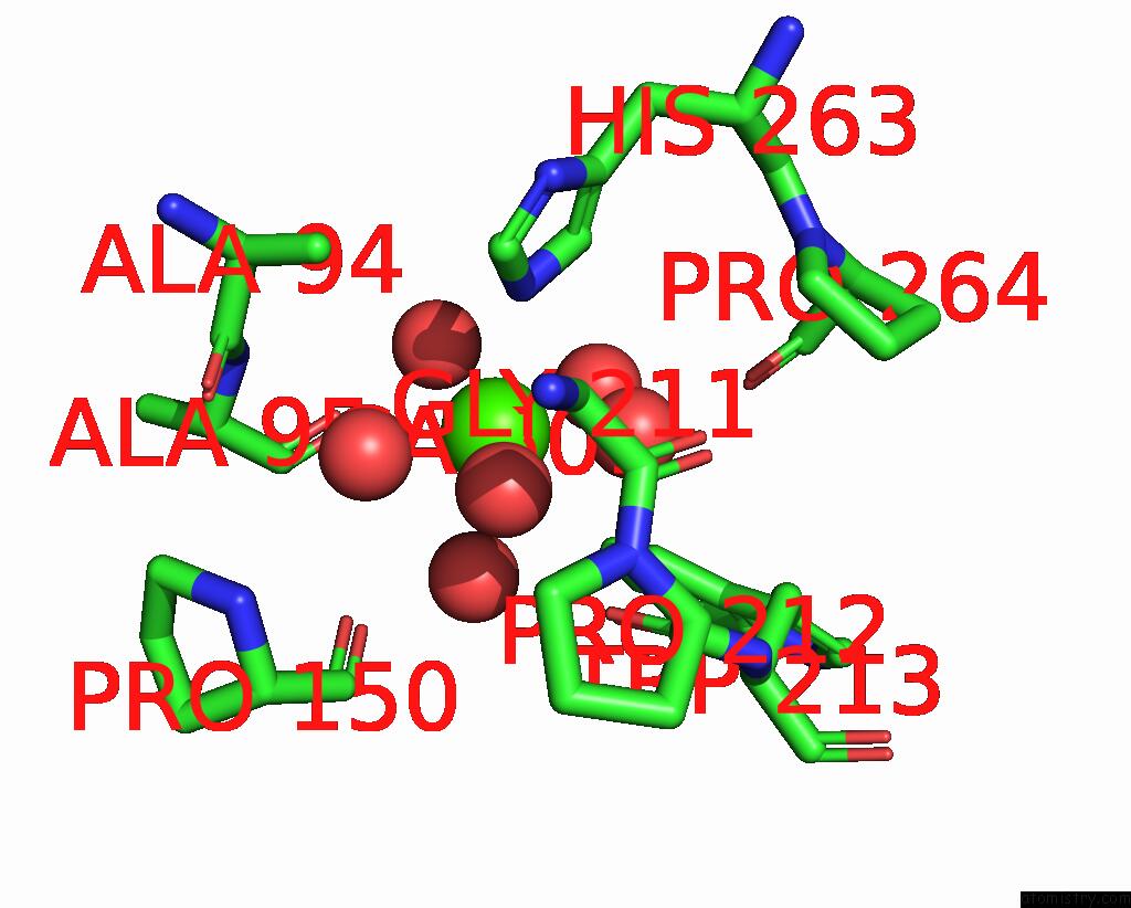
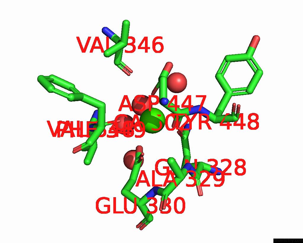
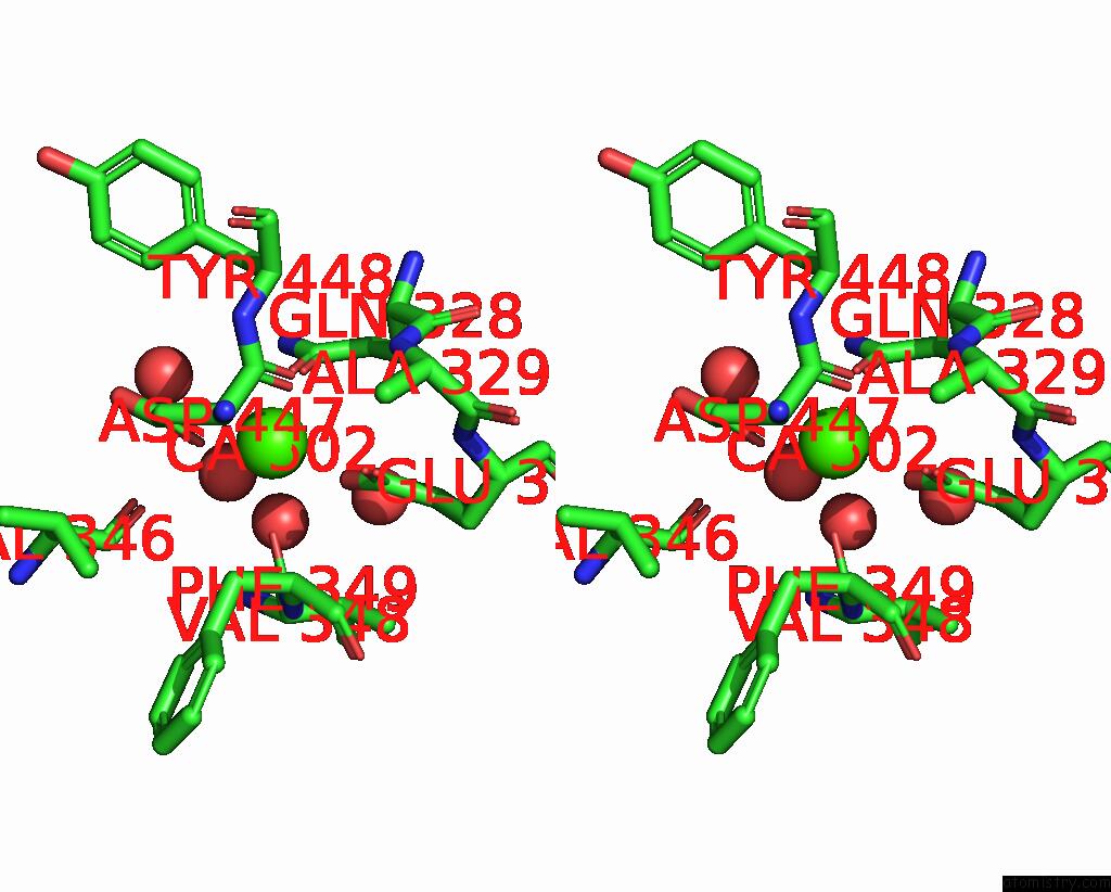
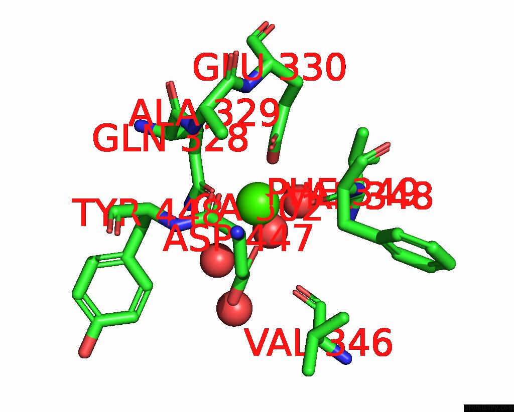
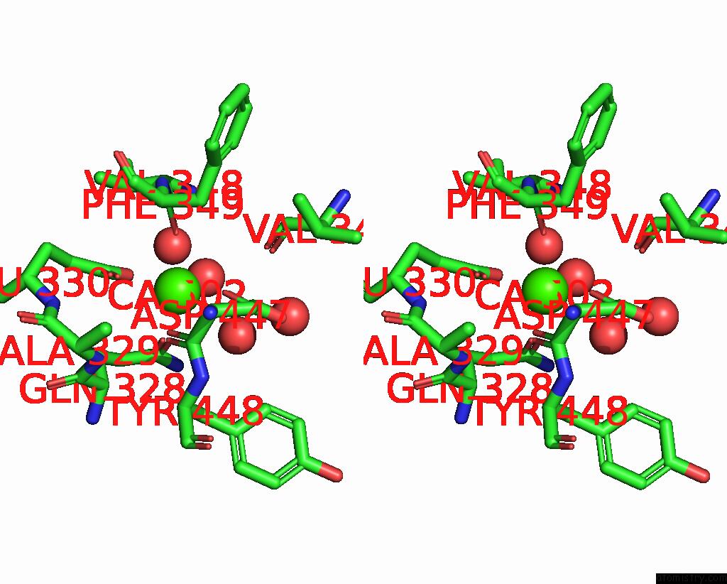
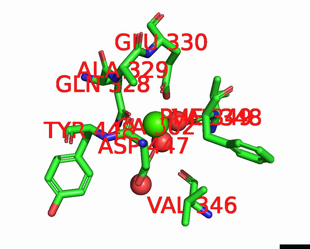
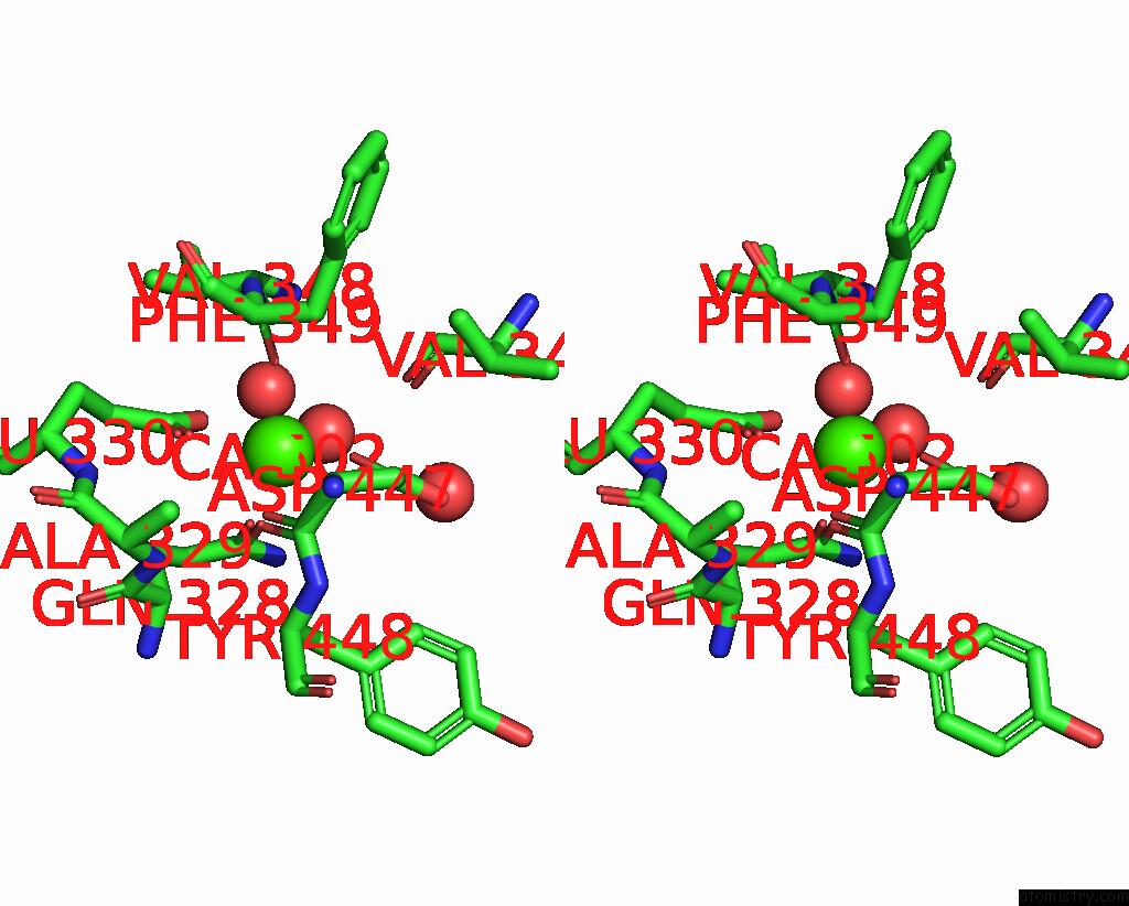
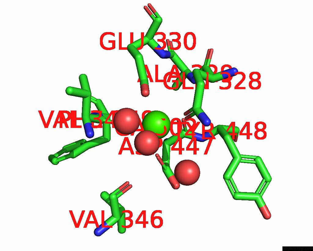
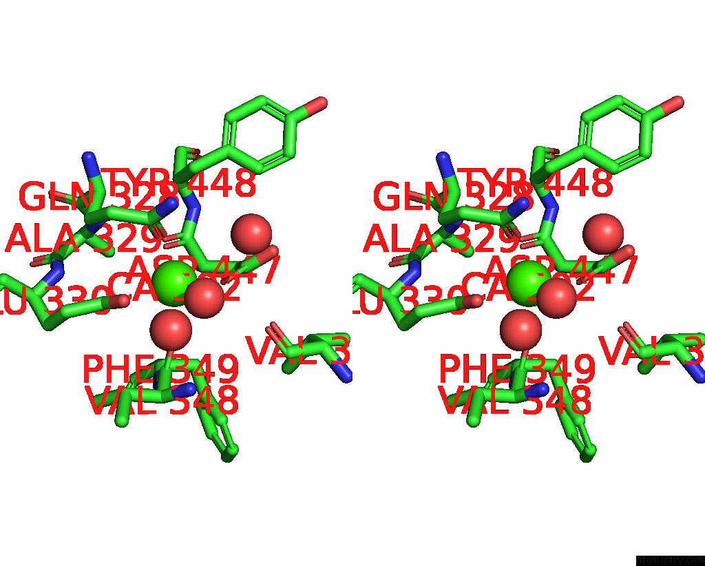
Calcium binding site 1 out of 16 in 8hcj
Go back to
Calcium binding site 1 out
of 16 in the Structure of GH43 Family Enzyme, Xylan 1, 4 Beta- Xylosidase From Pseudopedobacter Saltans
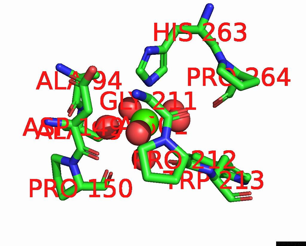
Mono view

Stereo pair view
Mono view
Stereo pair view
A full contact list of Calcium with other atoms in the Ca binding
site number 1 of Structure of GH43 Family Enzyme, Xylan 1, 4 Beta- Xylosidase From Pseudopedobacter Saltans within 5.0Å range:
|
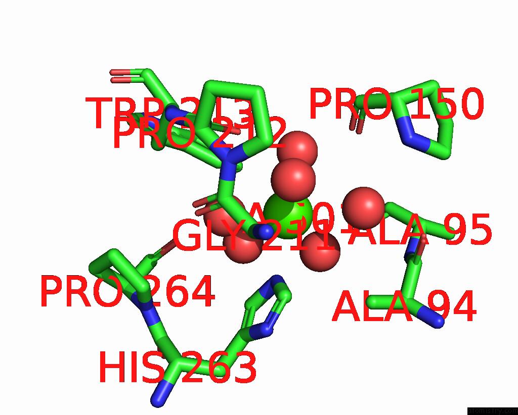
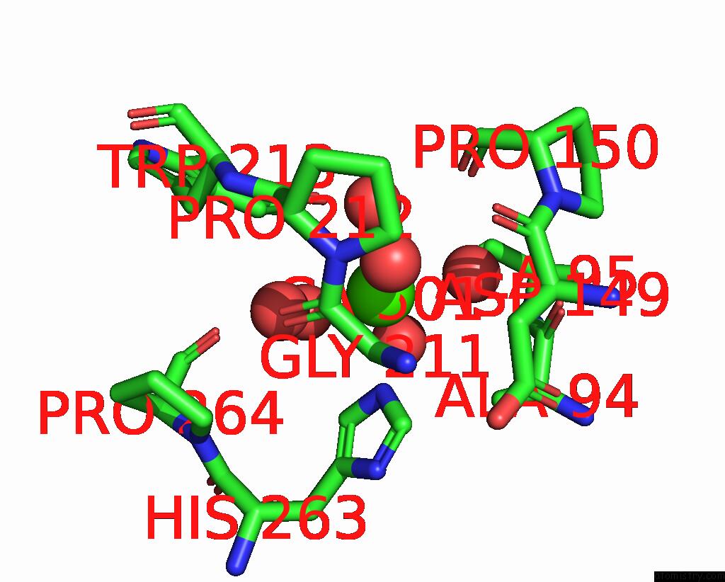
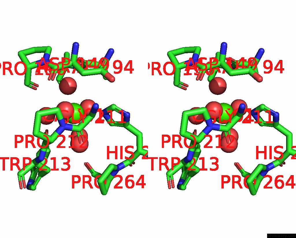
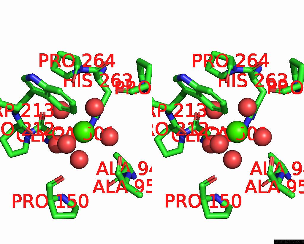
Calcium binding site 2 out of 16 in 8hcj
Go back to
Calcium binding site 2 out
of 16 in the Structure of GH43 Family Enzyme, Xylan 1, 4 Beta- Xylosidase From Pseudopedobacter Saltans
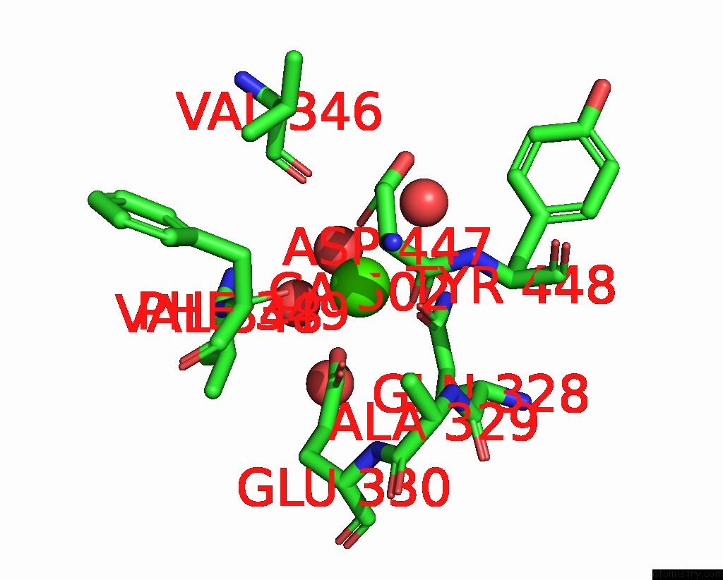
Mono view
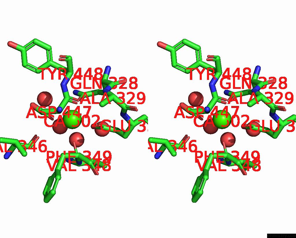
Stereo pair view
Mono view
Stereo pair view
A full contact list of Calcium with other atoms in the Ca binding
site number 2 of Structure of GH43 Family Enzyme, Xylan 1, 4 Beta- Xylosidase From Pseudopedobacter Saltans within 5.0Å range:
|
Calcium binding site 3 out of 16 in 8hcj
Go back to
Calcium binding site 3 out
of 16 in the Structure of GH43 Family Enzyme, Xylan 1, 4 Beta- Xylosidase From Pseudopedobacter Saltans
Mono view

Stereo pair view
Mono view
Stereo pair view
A full contact list of Calcium with other atoms in the Ca binding
site number 3 of Structure of GH43 Family Enzyme, Xylan 1, 4 Beta- Xylosidase From Pseudopedobacter Saltans within 5.0Å range:
|
Calcium binding site 4 out of 16 in 8hcj
Go back to
Calcium binding site 4 out
of 16 in the Structure of GH43 Family Enzyme, Xylan 1, 4 Beta- Xylosidase From Pseudopedobacter Saltans
Mono view
Stereo pair view
Mono view
Stereo pair view
A full contact list of Calcium with other atoms in the Ca binding
site number 4 of Structure of GH43 Family Enzyme, Xylan 1, 4 Beta- Xylosidase From Pseudopedobacter Saltans within 5.0Å range:
|
Calcium binding site 5 out of 16 in 8hcj
Go back to
Calcium binding site 5 out
of 16 in the Structure of GH43 Family Enzyme, Xylan 1, 4 Beta- Xylosidase From Pseudopedobacter Saltans
Mono view

Stereo pair view
Mono view
Stereo pair view
A full contact list of Calcium with other atoms in the Ca binding
site number 5 of Structure of GH43 Family Enzyme, Xylan 1, 4 Beta- Xylosidase From Pseudopedobacter Saltans within 5.0Å range:
|
Calcium binding site 6 out of 16 in 8hcj
Go back to
Calcium binding site 6 out
of 16 in the Structure of GH43 Family Enzyme, Xylan 1, 4 Beta- Xylosidase From Pseudopedobacter Saltans
Mono view
Stereo pair view
Mono view
Stereo pair view
A full contact list of Calcium with other atoms in the Ca binding
site number 6 of Structure of GH43 Family Enzyme, Xylan 1, 4 Beta- Xylosidase From Pseudopedobacter Saltans within 5.0Å range:
|
Calcium binding site 7 out of 16 in 8hcj
Go back to
Calcium binding site 7 out
of 16 in the Structure of GH43 Family Enzyme, Xylan 1, 4 Beta- Xylosidase From Pseudopedobacter Saltans
Mono view
Stereo pair view
Mono view
Stereo pair view
A full contact list of Calcium with other atoms in the Ca binding
site number 7 of Structure of GH43 Family Enzyme, Xylan 1, 4 Beta- Xylosidase From Pseudopedobacter Saltans within 5.0Å range:
|
Calcium binding site 8 out of 16 in 8hcj
Go back to
Calcium binding site 8 out
of 16 in the Structure of GH43 Family Enzyme, Xylan 1, 4 Beta- Xylosidase From Pseudopedobacter Saltans
Mono view
Stereo pair view
Mono view
Stereo pair view
A full contact list of Calcium with other atoms in the Ca binding
site number 8 of Structure of GH43 Family Enzyme, Xylan 1, 4 Beta- Xylosidase From Pseudopedobacter Saltans within 5.0Å range:
|
Calcium binding site 9 out of 16 in 8hcj
Go back to
Calcium binding site 9 out
of 16 in the Structure of GH43 Family Enzyme, Xylan 1, 4 Beta- Xylosidase From Pseudopedobacter Saltans

Mono view
Stereo pair view
Mono view
Stereo pair view
A full contact list of Calcium with other atoms in the Ca binding
site number 9 of Structure of GH43 Family Enzyme, Xylan 1, 4 Beta- Xylosidase From Pseudopedobacter Saltans within 5.0Å range:
|
Calcium binding site 10 out of 16 in 8hcj
Go back to
Calcium binding site 10 out
of 16 in the Structure of GH43 Family Enzyme, Xylan 1, 4 Beta- Xylosidase From Pseudopedobacter Saltans
Mono view
Stereo pair view
Mono view
Stereo pair view
A full contact list of Calcium with other atoms in the Ca binding
site number 10 of Structure of GH43 Family Enzyme, Xylan 1, 4 Beta- Xylosidase From Pseudopedobacter Saltans within 5.0Å range:
|
Reference:
P.Vishwakarma,
P.Kaur,
E.Sachdeva,
A.S.Ethayathulla,
U.Das,
A.Goyal.
Structure of GH43 Family Enzyme, Xylan 1, 4 Beta- Xylosidase From Pseudopedobacter Saltans To Be Published.
Page generated: Fri Jul 19 09:25:03 2024
Last articles
Zn in 9JYWZn in 9IR4
Zn in 9IR3
Zn in 9GMX
Zn in 9GMW
Zn in 9JEJ
Zn in 9ERF
Zn in 9ERE
Zn in 9EGV
Zn in 9EGW