Calcium in PDB 8qod: Crystal Structure of Fviia in Complex with A Benzamidine-Based Inhibitor
Enzymatic activity of Crystal Structure of Fviia in Complex with A Benzamidine-Based Inhibitor
All present enzymatic activity of Crystal Structure of Fviia in Complex with A Benzamidine-Based Inhibitor:
3.4.21.21;
3.4.21.21;
Protein crystallography data
The structure of Crystal Structure of Fviia in Complex with A Benzamidine-Based Inhibitor, PDB code: 8qod
was solved by
H.A.Schreuder,
A.Liesum,
with X-Ray Crystallography technique. A brief refinement statistics is given in the table below:
| Resolution Low / High (Å) | 22.88 / 1.57 |
| Space group | P 21 21 21 |
| Cell size a, b, c (Å), α, β, γ (°) | 69.811, 81.51, 125.498, 90, 90, 90 |
| R / Rfree (%) | 18.2 / 21.4 |
Other elements in 8qod:
The structure of Crystal Structure of Fviia in Complex with A Benzamidine-Based Inhibitor also contains other interesting chemical elements:
| Bromine | (Br) | 1 atom |
| Chlorine | (Cl) | 2 atoms |
| Magnesium | (Mg) | 1 atom |
Calcium Binding Sites:
The binding sites of Calcium atom in the Crystal Structure of Fviia in Complex with A Benzamidine-Based Inhibitor
(pdb code 8qod). This binding sites where shown within
5.0 Angstroms radius around Calcium atom.
In total 4 binding sites of Calcium where determined in the Crystal Structure of Fviia in Complex with A Benzamidine-Based Inhibitor, PDB code: 8qod:
Jump to Calcium binding site number: 1; 2; 3; 4;
In total 4 binding sites of Calcium where determined in the Crystal Structure of Fviia in Complex with A Benzamidine-Based Inhibitor, PDB code: 8qod:
Jump to Calcium binding site number: 1; 2; 3; 4;
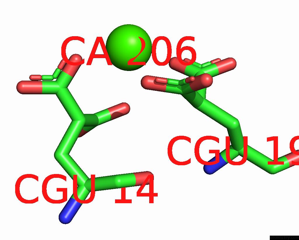
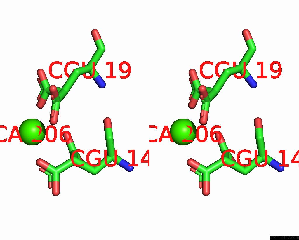
Calcium binding site 1 out of 4 in 8qod
Go back to
Calcium binding site 1 out
of 4 in the Crystal Structure of Fviia in Complex with A Benzamidine-Based Inhibitor
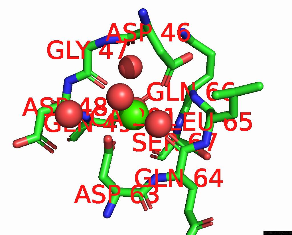
Mono view
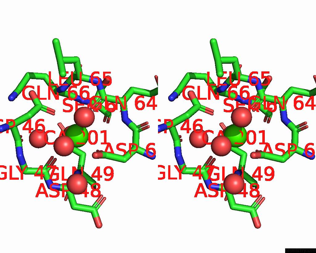
Stereo pair view
Mono view
Stereo pair view
A full contact list of Calcium with other atoms in the Ca binding
site number 1 of Crystal Structure of Fviia in Complex with A Benzamidine-Based Inhibitor within 5.0Å range:
|
Calcium binding site 2 out of 4 in 8qod
Go back to
Calcium binding site 2 out
of 4 in the Crystal Structure of Fviia in Complex with A Benzamidine-Based Inhibitor
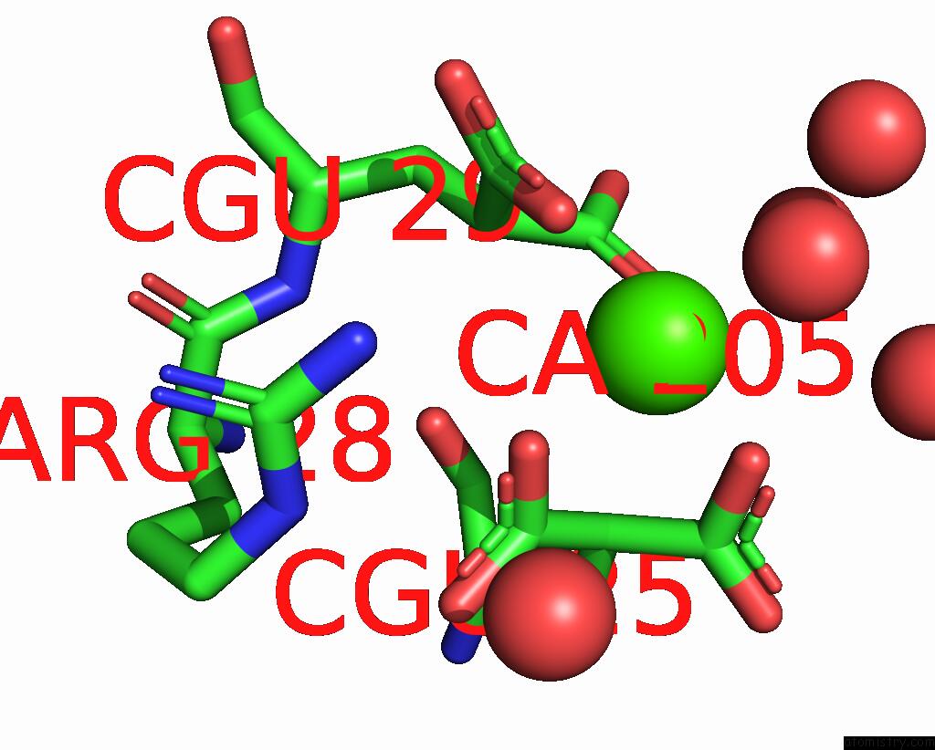
Mono view
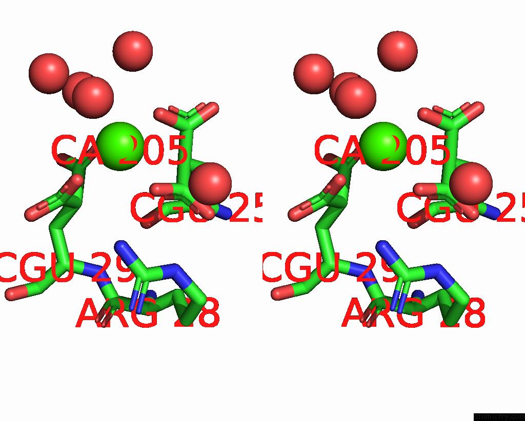
Stereo pair view
Mono view
Stereo pair view
A full contact list of Calcium with other atoms in the Ca binding
site number 2 of Crystal Structure of Fviia in Complex with A Benzamidine-Based Inhibitor within 5.0Å range:
|
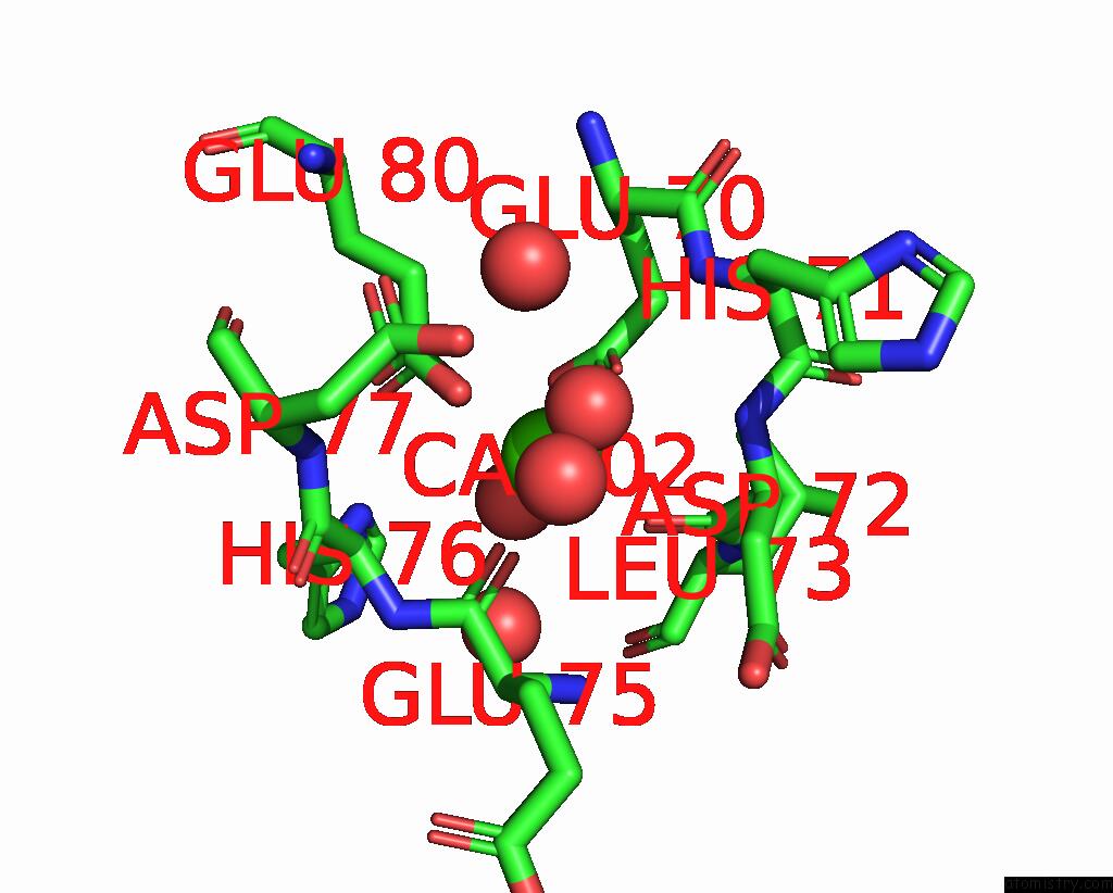
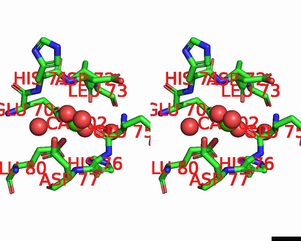
Calcium binding site 3 out of 4 in 8qod
Go back to
Calcium binding site 3 out
of 4 in the Crystal Structure of Fviia in Complex with A Benzamidine-Based Inhibitor
Mono view
Stereo pair view
Mono view
Stereo pair view
A full contact list of Calcium with other atoms in the Ca binding
site number 3 of Crystal Structure of Fviia in Complex with A Benzamidine-Based Inhibitor within 5.0Å range:
|
Calcium binding site 4 out of 4 in 8qod
Go back to
Calcium binding site 4 out
of 4 in the Crystal Structure of Fviia in Complex with A Benzamidine-Based Inhibitor
Mono view
Stereo pair view
Mono view
Stereo pair view
A full contact list of Calcium with other atoms in the Ca binding
site number 4 of Crystal Structure of Fviia in Complex with A Benzamidine-Based Inhibitor within 5.0Å range:
|
Reference:
L.Tesmer,
H.Hans Matter,
O.Klingler,
M.Schudok,
G.Hessler,
A.R.Mehdipour,
S.Guessregen,
G.Hummer,
H.A.Schreuder.
Crystallography and Molecular Simulations Capture S1 Pocket Collapse in Allosteric Regulation of Factor Viia and Other Serine Proteases. To Be Published.
Page generated: Thu Oct 31 17:11:19 2024
Last articles
Zn in 9J0NZn in 9J0O
Zn in 9J0P
Zn in 9FJX
Zn in 9EKB
Zn in 9C0F
Zn in 9CAH
Zn in 9CH0
Zn in 9CH3
Zn in 9CH1