Calcium in PDB 8s3c: Crystal Structure of Medicago Truncatula Glutamate Dehydrogenase 2 (Unliganded)
Protein crystallography data
The structure of Crystal Structure of Medicago Truncatula Glutamate Dehydrogenase 2 (Unliganded), PDB code: 8s3c
was solved by
M.Grzechowiak,
M.Ruszkowski,
with X-Ray Crystallography technique. A brief refinement statistics is given in the table below:
| Resolution Low / High (Å) | 47.83 / 2.20 |
| Space group | C 2 2 21 |
| Cell size a, b, c (Å), α, β, γ (°) | 95.651, 164.554, 195.412, 90, 90, 90 |
| R / Rfree (%) | 17.4 / 20.4 |
Other elements in 8s3c:
The structure of Crystal Structure of Medicago Truncatula Glutamate Dehydrogenase 2 (Unliganded) also contains other interesting chemical elements:
| Sodium | (Na) | 2 atoms |
Calcium Binding Sites:
The binding sites of Calcium atom in the Crystal Structure of Medicago Truncatula Glutamate Dehydrogenase 2 (Unliganded)
(pdb code 8s3c). This binding sites where shown within
5.0 Angstroms radius around Calcium atom.
In total 3 binding sites of Calcium where determined in the Crystal Structure of Medicago Truncatula Glutamate Dehydrogenase 2 (Unliganded), PDB code: 8s3c:
Jump to Calcium binding site number: 1; 2; 3;
In total 3 binding sites of Calcium where determined in the Crystal Structure of Medicago Truncatula Glutamate Dehydrogenase 2 (Unliganded), PDB code: 8s3c:
Jump to Calcium binding site number: 1; 2; 3;
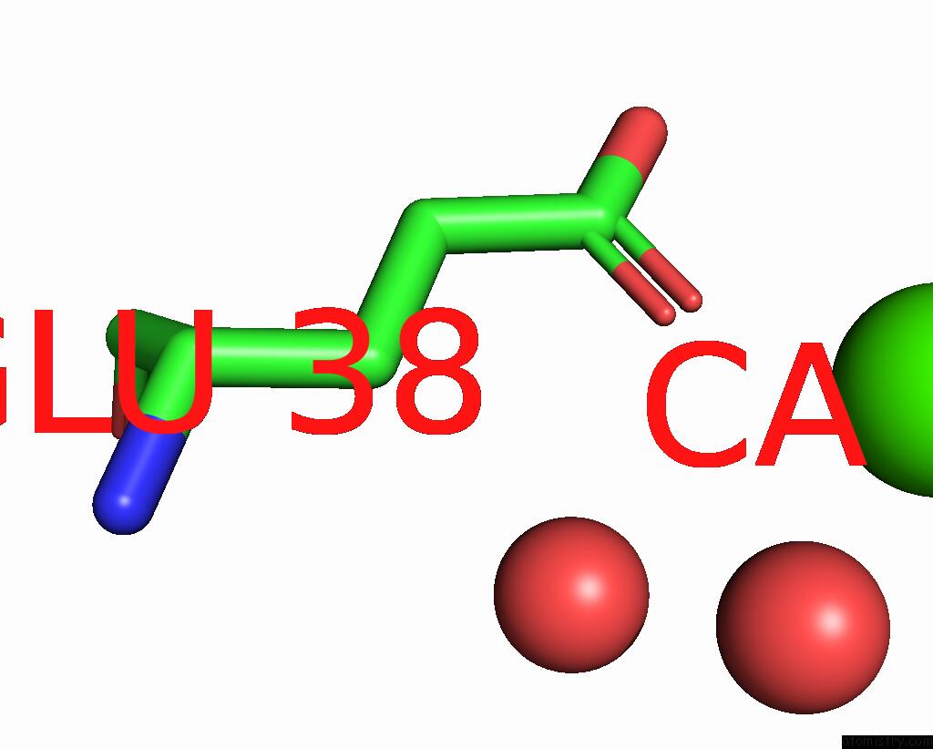
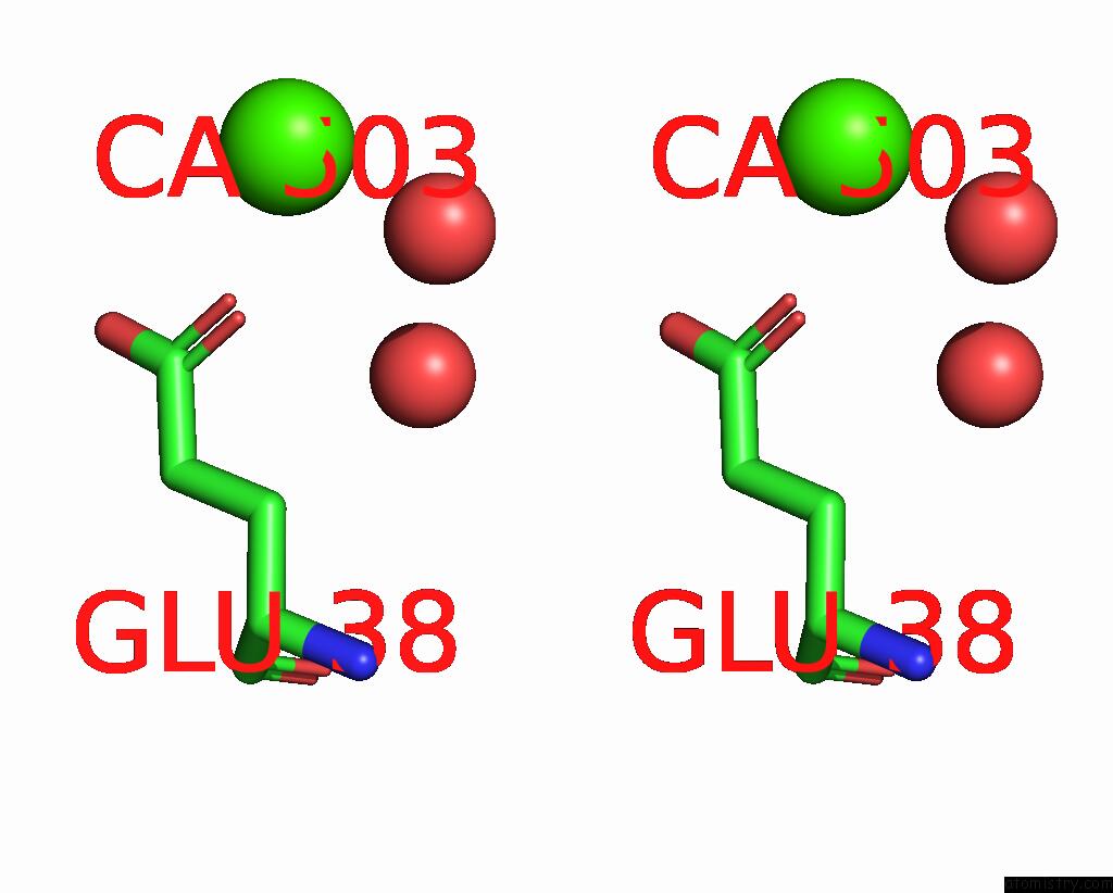
Calcium binding site 1 out of 3 in 8s3c
Go back to
Calcium binding site 1 out
of 3 in the Crystal Structure of Medicago Truncatula Glutamate Dehydrogenase 2 (Unliganded)
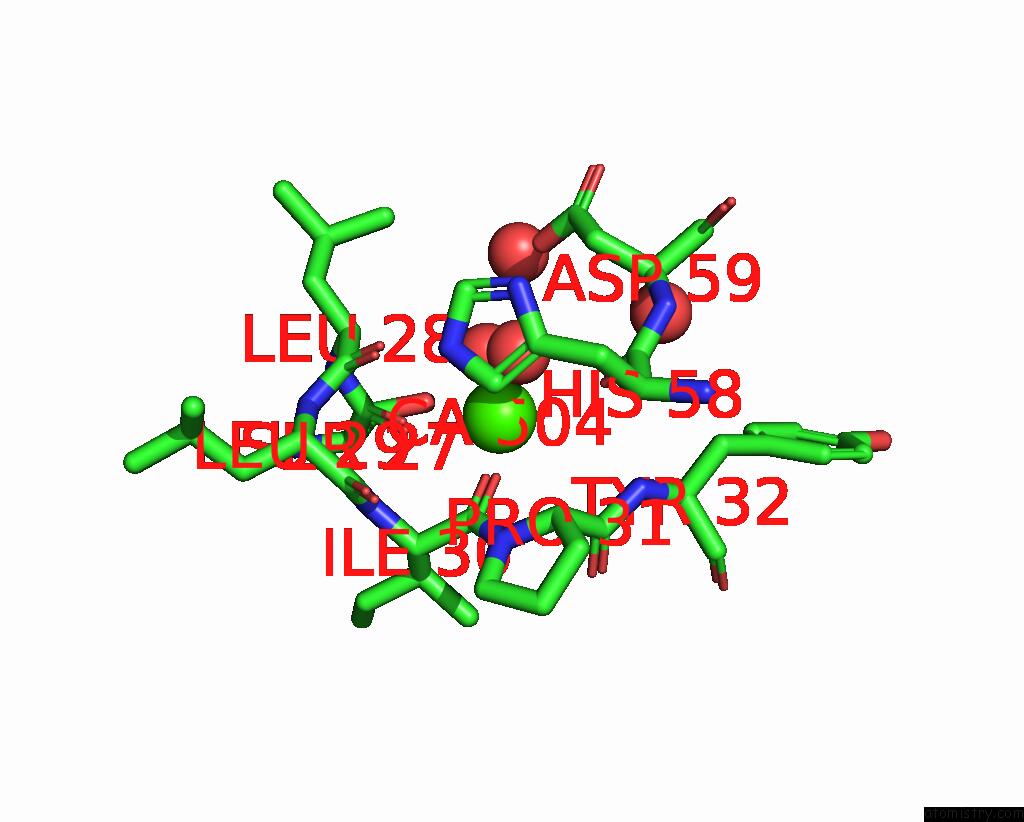
Mono view
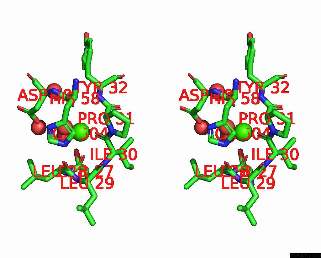
Stereo pair view
Mono view
Stereo pair view
A full contact list of Calcium with other atoms in the Ca binding
site number 1 of Crystal Structure of Medicago Truncatula Glutamate Dehydrogenase 2 (Unliganded) within 5.0Å range:
|
Calcium binding site 2 out of 3 in 8s3c
Go back to
Calcium binding site 2 out
of 3 in the Crystal Structure of Medicago Truncatula Glutamate Dehydrogenase 2 (Unliganded)
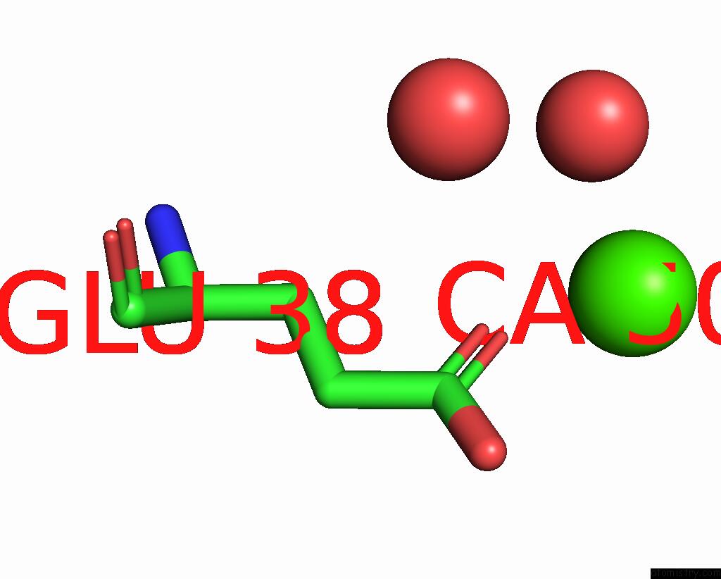
Mono view
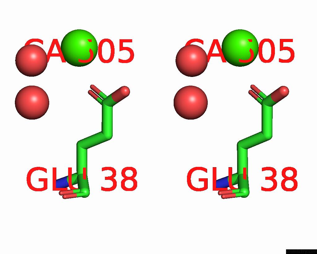
Stereo pair view
Mono view
Stereo pair view
A full contact list of Calcium with other atoms in the Ca binding
site number 2 of Crystal Structure of Medicago Truncatula Glutamate Dehydrogenase 2 (Unliganded) within 5.0Å range:
|
Calcium binding site 3 out of 3 in 8s3c
Go back to
Calcium binding site 3 out
of 3 in the Crystal Structure of Medicago Truncatula Glutamate Dehydrogenase 2 (Unliganded)
Mono view
Stereo pair view
Mono view
Stereo pair view
A full contact list of Calcium with other atoms in the Ca binding
site number 3 of Crystal Structure of Medicago Truncatula Glutamate Dehydrogenase 2 (Unliganded) within 5.0Å range:
|
Reference:
M.Grzechowiak,
J.Sliwiak,
A.Link,
M.Ruszkowski.
Legume-Type Glutamate Dehydrogenase: Structure, Activity, and Inhibition Studies. Int.J.Biol.Macromol. V. 278 34648 2024.
ISSN: ISSN 0141-8130
PubMed: 39142482
DOI: 10.1016/J.IJBIOMAC.2024.134648
Page generated: Sat Sep 28 09:02:56 2024
ISSN: ISSN 0141-8130
PubMed: 39142482
DOI: 10.1016/J.IJBIOMAC.2024.134648
Last articles
Zn in 9JYWZn in 9IR4
Zn in 9IR3
Zn in 9GMX
Zn in 9GMW
Zn in 9JEJ
Zn in 9ERF
Zn in 9ERE
Zn in 9EGV
Zn in 9EGW