Calcium in PDB 8s3d: Crystal Structure of Medicago Truncatula Glutamate Dehydrogenase 2 in Complex with 2-Amino-2-Hydroxyglutarate (Reaction Intermediate) and Nad
Protein crystallography data
The structure of Crystal Structure of Medicago Truncatula Glutamate Dehydrogenase 2 in Complex with 2-Amino-2-Hydroxyglutarate (Reaction Intermediate) and Nad, PDB code: 8s3d
was solved by
M.Grzechowiak,
M.Ruszkowski,
with X-Ray Crystallography technique. A brief refinement statistics is given in the table below:
Resolution Low / High (Å)
|
65.89 /
1.65
|
Space group
|
P 21 21 21
|
Cell size a, b, c (Å), α, β, γ (°)
|
111.832,
155.707,
163.089,
90,
90,
90
|
R / Rfree (%)
|
14.7 /
17.4
|
Other elements in 8s3d:
The structure of Crystal Structure of Medicago Truncatula Glutamate Dehydrogenase 2 in Complex with 2-Amino-2-Hydroxyglutarate (Reaction Intermediate) and Nad also contains other interesting chemical elements:
Calcium Binding Sites:
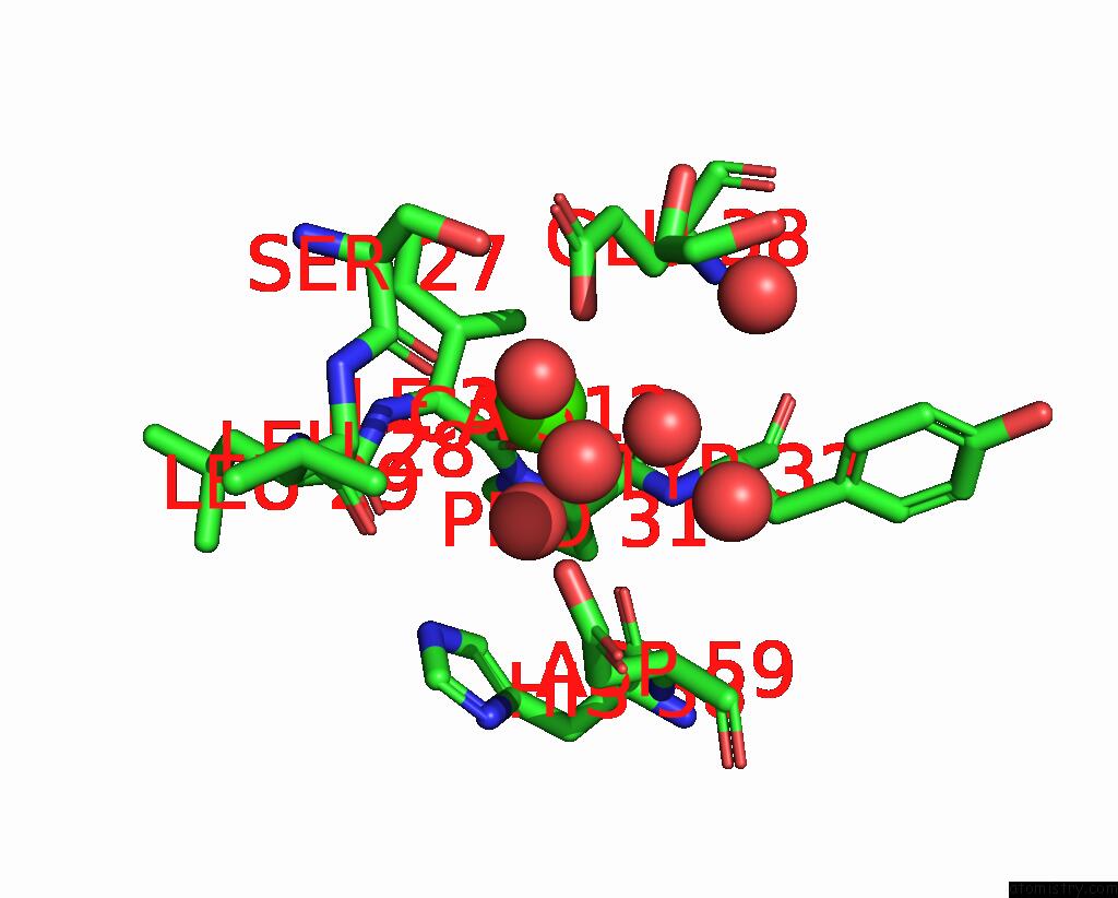
The binding sites of Calcium atom in the Crystal Structure of Medicago Truncatula Glutamate Dehydrogenase 2 in Complex with 2-Amino-2-Hydroxyglutarate (Reaction Intermediate) and Nad
(pdb code 8s3d). This binding sites where shown within
5.0 Angstroms radius around Calcium atom.
In total 6 binding sites of Calcium where determined in the
Crystal Structure of Medicago Truncatula Glutamate Dehydrogenase 2 in Complex with 2-Amino-2-Hydroxyglutarate (Reaction Intermediate) and Nad, PDB code: 8s3d:
Jump to Calcium binding site number:
1;
2;
3;
4;
5;
6;
Calcium binding site 1 out
of 6 in 8s3d
Go back to
Calcium Binding Sites List in 8s3d
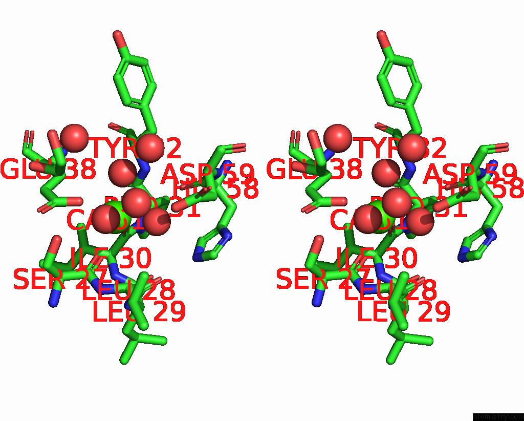
Calcium binding site 1 out
of 6 in the Crystal Structure of Medicago Truncatula Glutamate Dehydrogenase 2 in Complex with 2-Amino-2-Hydroxyglutarate (Reaction Intermediate) and Nad
Mono view
Stereo pair view
|
A full contact list of Calcium with other atoms in the Ca binding
site number 1 of Crystal Structure of Medicago Truncatula Glutamate Dehydrogenase 2 in Complex with 2-Amino-2-Hydroxyglutarate (Reaction Intermediate) and Nad within 5.0Å range:
probe
|
atom
|
residue
|
distance (Å)
|
B
|
Occ
|
A:Ca512
b:19.6
occ:1.00
|
OE2
|
E:GLU38
|
2.2
|
19.9
|
1.0
|
O
|
A:ILE30
|
2.3
|
18.4
|
1.0
|
O
|
A:HOH645
|
2.3
|
24.1
|
1.0
|
O
|
A:SER27
|
2.3
|
20.5
|
1.0
|
O
|
A:HOH769
|
2.4
|
20.9
|
1.0
|
O
|
E:HOH891
|
2.4
|
28.6
|
1.0
|
CD
|
E:GLU38
|
3.4
|
18.9
|
1.0
|
C
|
A:SER27
|
3.5
|
18.4
|
1.0
|
C
|
A:ILE30
|
3.5
|
18.6
|
1.0
|
OE1
|
E:GLU38
|
4.0
|
20.3
|
1.0
|
OG
|
A:SER27
|
4.0
|
18.9
|
1.0
|
CA
|
A:LEU28
|
4.2
|
17.7
|
1.0
|
O
|
A:LEU28
|
4.2
|
19.9
|
1.0
|
C
|
A:LEU28
|
4.2
|
20.6
|
1.0
|
O
|
A:HIS58
|
4.2
|
16.4
|
1.0
|
N
|
A:LEU28
|
4.3
|
17.6
|
1.0
|
N
|
A:TYR32
|
4.3
|
16.3
|
1.0
|
CA
|
A:ILE30
|
4.3
|
17.9
|
1.0
|
N
|
A:ILE30
|
4.3
|
19.3
|
1.0
|
C
|
A:PRO31
|
4.3
|
15.5
|
1.0
|
OD2
|
A:ASP59
|
4.4
|
25.4
|
1.0
|
CB
|
A:ILE30
|
4.4
|
17.8
|
1.0
|
CA
|
A:SER27
|
4.4
|
18.3
|
1.0
|
N
|
A:PRO31
|
4.5
|
19.5
|
1.0
|
CG
|
E:GLU38
|
4.6
|
17.3
|
1.0
|
CA
|
A:PRO31
|
4.6
|
17.0
|
1.0
|
O
|
A:HOH894
|
4.6
|
22.8
|
1.0
|
CB
|
E:GLU38
|
4.7
|
16.0
|
1.0
|
O
|
A:HOH644
|
4.8
|
19.9
|
1.0
|
O
|
A:PRO31
|
4.8
|
18.1
|
1.0
|
O
|
E:HOH818
|
4.8
|
29.8
|
1.0
|
CB
|
A:SER27
|
4.8
|
22.1
|
1.0
|
CA
|
A:TYR32
|
4.9
|
15.4
|
1.0
|
N
|
A:LEU29
|
4.9
|
18.8
|
1.0
|
C
|
A:LEU29
|
4.9
|
23.2
|
1.0
|
C1
|
A:EDO507
|
5.0
|
37.4
|
1.0
|
CG2
|
A:ILE30
|
5.0
|
19.3
|
1.0
|
|
Calcium binding site 2 out
of 6 in 8s3d
Go back to
Calcium Binding Sites List in 8s3d
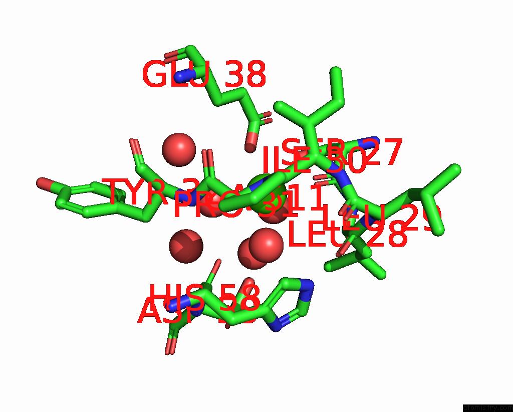
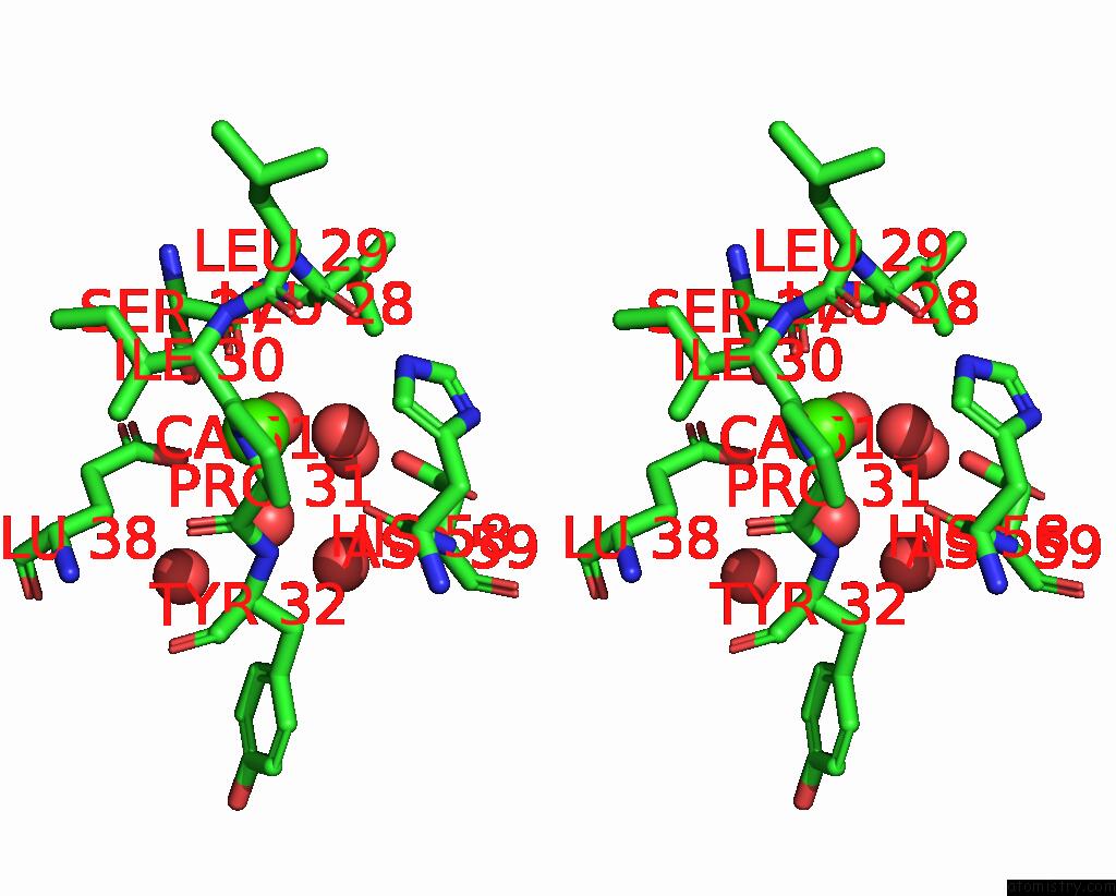
Calcium binding site 2 out
of 6 in the Crystal Structure of Medicago Truncatula Glutamate Dehydrogenase 2 in Complex with 2-Amino-2-Hydroxyglutarate (Reaction Intermediate) and Nad
Mono view
Stereo pair view
|
A full contact list of Calcium with other atoms in the Ca binding
site number 2 of Crystal Structure of Medicago Truncatula Glutamate Dehydrogenase 2 in Complex with 2-Amino-2-Hydroxyglutarate (Reaction Intermediate) and Nad within 5.0Å range:
probe
|
atom
|
residue
|
distance (Å)
|
B
|
Occ
|
B:Ca511
b:18.3
occ:1.00
|
OE2
|
D:GLU38
|
2.3
|
18.6
|
1.0
|
O
|
B:HOH712
|
2.3
|
22.2
|
1.0
|
O
|
B:SER27
|
2.3
|
18.6
|
1.0
|
O
|
B:HOH788
|
2.3
|
18.9
|
1.0
|
O
|
B:ILE30
|
2.3
|
19.5
|
1.0
|
O
|
D:HOH796
|
2.4
|
25.2
|
1.0
|
CD
|
D:GLU38
|
3.4
|
20.1
|
1.0
|
C
|
B:SER27
|
3.4
|
18.0
|
1.0
|
C
|
B:ILE30
|
3.6
|
19.2
|
1.0
|
OG
|
B:SER27
|
4.0
|
19.5
|
1.0
|
OE1
|
D:GLU38
|
4.0
|
19.1
|
1.0
|
CA
|
B:LEU28
|
4.1
|
19.4
|
1.0
|
N
|
B:LEU28
|
4.2
|
18.0
|
1.0
|
N
|
B:ILE30
|
4.2
|
20.0
|
1.0
|
C
|
B:LEU28
|
4.2
|
20.2
|
1.0
|
O
|
B:LEU28
|
4.3
|
21.3
|
1.0
|
CA
|
B:ILE30
|
4.3
|
19.5
|
1.0
|
O
|
B:HIS58
|
4.3
|
16.5
|
1.0
|
N
|
B:TYR32
|
4.3
|
16.0
|
1.0
|
C
|
B:PRO31
|
4.4
|
15.9
|
1.0
|
OD2
|
B:ASP59
|
4.4
|
23.5
|
1.0
|
CA
|
B:SER27
|
4.4
|
19.0
|
1.0
|
CB
|
B:ILE30
|
4.4
|
17.7
|
1.0
|
N
|
B:PRO31
|
4.6
|
18.1
|
1.0
|
O
|
B:HOH892
|
4.6
|
20.8
|
1.0
|
CG
|
D:GLU38
|
4.6
|
17.3
|
1.0
|
CA
|
B:PRO31
|
4.6
|
15.4
|
1.0
|
CB
|
D:GLU38
|
4.7
|
20.2
|
1.0
|
O
|
B:HOH618
|
4.7
|
19.0
|
1.0
|
O
|
D:HOH610
|
4.8
|
25.4
|
1.0
|
O
|
B:PRO31
|
4.8
|
16.5
|
1.0
|
CB
|
B:SER27
|
4.8
|
19.7
|
1.0
|
N
|
B:LEU29
|
4.9
|
19.9
|
1.0
|
C
|
B:LEU29
|
4.9
|
23.6
|
1.0
|
CA
|
B:TYR32
|
4.9
|
16.6
|
1.0
|
|
Calcium binding site 3 out
of 6 in 8s3d
Go back to
Calcium Binding Sites List in 8s3d
Calcium binding site 3 out
of 6 in the Crystal Structure of Medicago Truncatula Glutamate Dehydrogenase 2 in Complex with 2-Amino-2-Hydroxyglutarate (Reaction Intermediate) and Nad
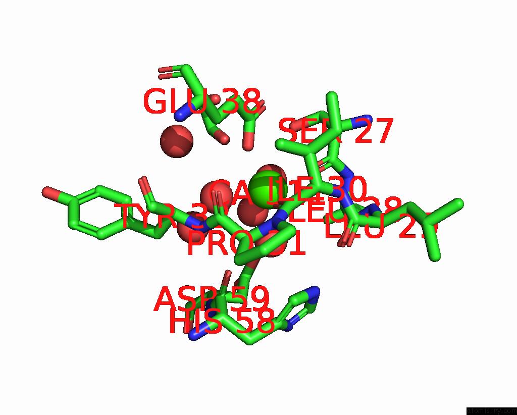
Mono view
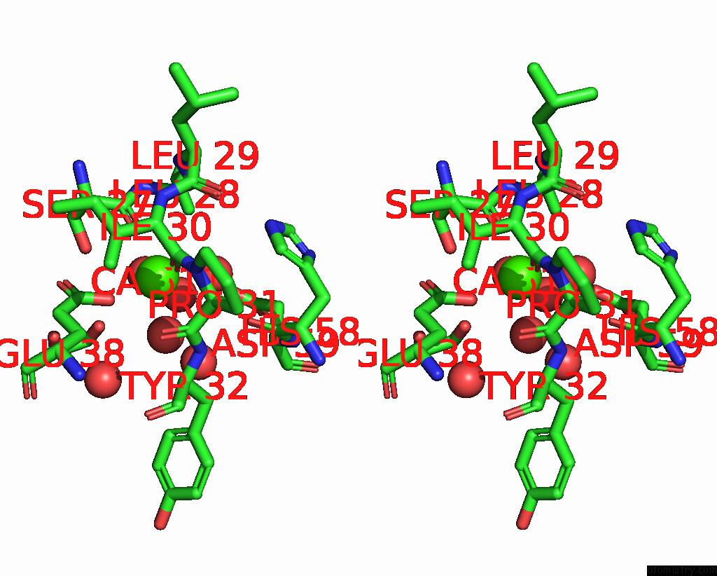
Stereo pair view
|
A full contact list of Calcium with other atoms in the Ca binding
site number 3 of Crystal Structure of Medicago Truncatula Glutamate Dehydrogenase 2 in Complex with 2-Amino-2-Hydroxyglutarate (Reaction Intermediate) and Nad within 5.0Å range:
probe
|
atom
|
residue
|
distance (Å)
|
B
|
Occ
|
C:Ca511
b:19.0
occ:1.00
|
OE2
|
F:GLU38
|
2.3
|
20.9
|
1.0
|
O
|
C:SER27
|
2.3
|
17.4
|
1.0
|
O
|
C:ILE30
|
2.3
|
17.7
|
1.0
|
O
|
F:HOH2640
|
2.4
|
29.4
|
1.0
|
O
|
C:HOH767
|
2.5
|
18.4
|
1.0
|
O
|
C:HOH635
|
2.5
|
25.2
|
1.0
|
CD
|
F:GLU38
|
3.4
|
21.2
|
1.0
|
C
|
C:SER27
|
3.4
|
17.8
|
1.0
|
C
|
C:ILE30
|
3.5
|
19.2
|
1.0
|
OE1
|
F:GLU38
|
3.9
|
19.2
|
1.0
|
OG
|
C:SER27
|
4.0
|
20.4
|
1.0
|
CA
|
C:LEU28
|
4.1
|
18.5
|
1.0
|
N
|
C:LEU28
|
4.2
|
18.5
|
1.0
|
O
|
C:LEU28
|
4.2
|
20.1
|
1.0
|
N
|
C:ILE30
|
4.2
|
19.1
|
1.0
|
C
|
C:LEU28
|
4.2
|
17.7
|
1.0
|
CA
|
C:ILE30
|
4.2
|
17.2
|
1.0
|
O
|
C:HIS58
|
4.3
|
15.3
|
1.0
|
N
|
C:TYR32
|
4.3
|
16.1
|
1.0
|
CB
|
C:ILE30
|
4.3
|
19.1
|
1.0
|
C
|
C:PRO31
|
4.4
|
17.1
|
1.0
|
CA
|
C:SER27
|
4.4
|
18.5
|
1.0
|
OD2
|
C:ASP59
|
4.4
|
26.9
|
1.0
|
N
|
C:PRO31
|
4.5
|
18.3
|
1.0
|
CG
|
F:GLU38
|
4.6
|
20.2
|
1.0
|
O
|
C:HOH888
|
4.6
|
23.6
|
1.0
|
CA
|
C:PRO31
|
4.6
|
17.0
|
1.0
|
CB
|
F:GLU38
|
4.7
|
17.9
|
1.0
|
O
|
C:HOH625
|
4.7
|
20.2
|
1.0
|
O2
|
F:EDO2302
|
4.7
|
45.4
|
1.0
|
O
|
C:PRO31
|
4.8
|
18.6
|
1.0
|
O
|
F:HOH2628
|
4.9
|
26.9
|
1.0
|
CB
|
C:SER27
|
4.9
|
20.8
|
1.0
|
C
|
C:LEU29
|
4.9
|
24.4
|
1.0
|
N
|
C:LEU29
|
4.9
|
18.4
|
1.0
|
CG2
|
C:ILE30
|
4.9
|
21.1
|
1.0
|
CA
|
C:TYR32
|
4.9
|
17.4
|
1.0
|
|
Calcium binding site 4 out
of 6 in 8s3d
Go back to
Calcium Binding Sites List in 8s3d
Calcium binding site 4 out
of 6 in the Crystal Structure of Medicago Truncatula Glutamate Dehydrogenase 2 in Complex with 2-Amino-2-Hydroxyglutarate (Reaction Intermediate) and Nad
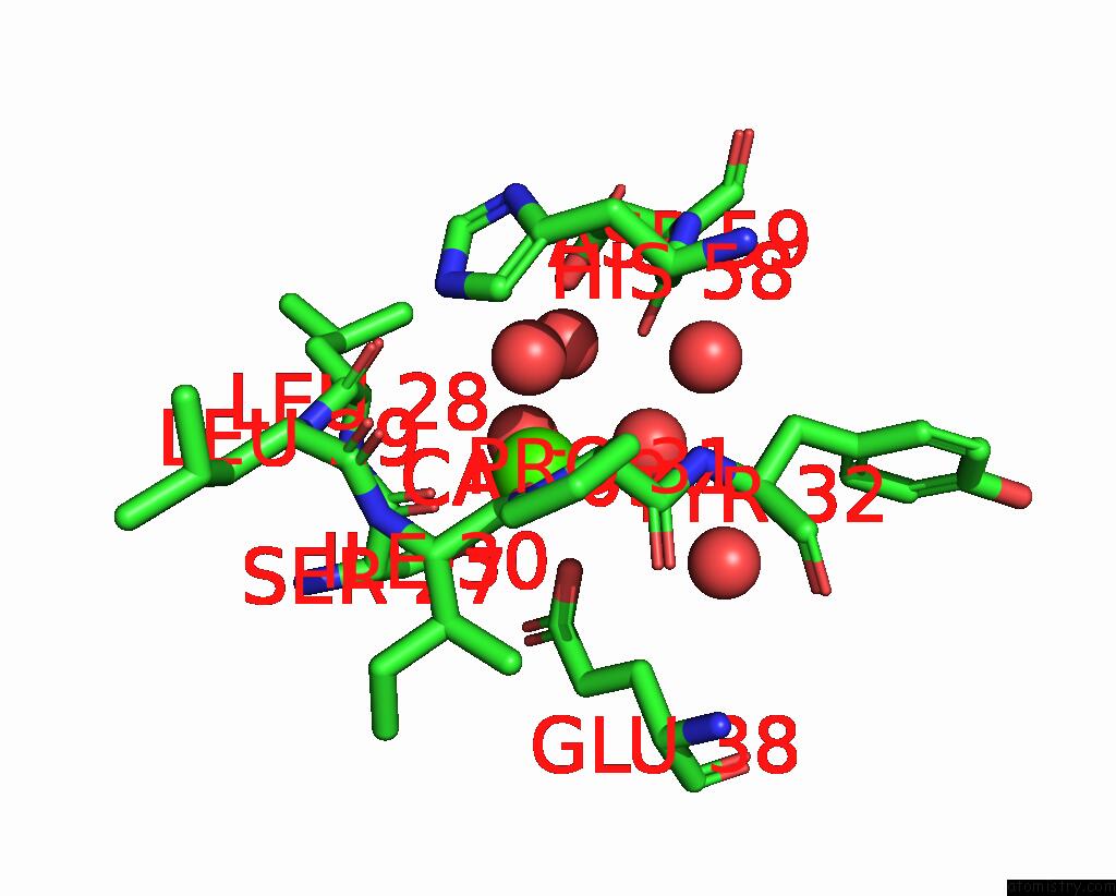
Mono view
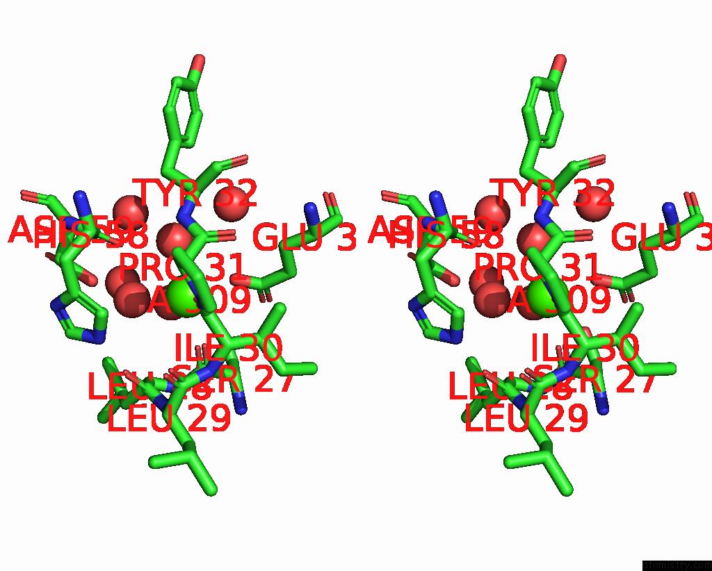
Stereo pair view
|
A full contact list of Calcium with other atoms in the Ca binding
site number 4 of Crystal Structure of Medicago Truncatula Glutamate Dehydrogenase 2 in Complex with 2-Amino-2-Hydroxyglutarate (Reaction Intermediate) and Nad within 5.0Å range:
probe
|
atom
|
residue
|
distance (Å)
|
B
|
Occ
|
D:Ca509
b:19.2
occ:1.00
|
OE2
|
B:GLU38
|
2.3
|
19.1
|
1.0
|
O
|
D:SER27
|
2.3
|
18.9
|
1.0
|
O
|
D:ILE30
|
2.3
|
19.4
|
1.0
|
O
|
D:HOH719
|
2.4
|
24.9
|
1.0
|
O
|
B:HOH889
|
2.4
|
28.5
|
1.0
|
O
|
D:HOH722
|
2.4
|
19.9
|
1.0
|
C
|
D:SER27
|
3.4
|
19.6
|
1.0
|
CD
|
B:GLU38
|
3.4
|
21.8
|
1.0
|
C
|
D:ILE30
|
3.5
|
18.0
|
1.0
|
OG
|
D:SER27
|
4.0
|
20.1
|
1.0
|
OE1
|
B:GLU38
|
4.0
|
20.3
|
1.0
|
CA
|
D:LEU28
|
4.1
|
22.0
|
1.0
|
N
|
D:LEU28
|
4.2
|
21.3
|
1.0
|
N
|
D:ILE30
|
4.2
|
18.8
|
1.0
|
C
|
D:LEU28
|
4.3
|
21.1
|
1.0
|
CA
|
D:ILE30
|
4.3
|
19.6
|
1.0
|
O
|
D:HIS58
|
4.3
|
19.1
|
1.0
|
O
|
D:LEU28
|
4.3
|
21.1
|
1.0
|
CA
|
D:SER27
|
4.4
|
19.9
|
1.0
|
OD2
|
D:ASP59
|
4.4
|
27.3
|
1.0
|
N
|
D:TYR32
|
4.4
|
16.8
|
1.0
|
C
|
D:PRO31
|
4.4
|
18.3
|
1.0
|
CB
|
D:ILE30
|
4.4
|
18.9
|
1.0
|
N
|
D:PRO31
|
4.5
|
20.5
|
1.0
|
CG
|
B:GLU38
|
4.5
|
18.2
|
1.0
|
O
|
D:HOH609
|
4.6
|
21.4
|
1.0
|
CA
|
D:PRO31
|
4.6
|
17.1
|
1.0
|
O
|
D:HOH857
|
4.6
|
25.2
|
1.0
|
CB
|
B:GLU38
|
4.7
|
17.4
|
1.0
|
O
|
B:HOH602
|
4.8
|
28.6
|
1.0
|
CB
|
D:SER27
|
4.8
|
20.1
|
1.0
|
O
|
D:PRO31
|
4.8
|
18.3
|
1.0
|
C
|
D:LEU29
|
4.9
|
22.8
|
1.0
|
N
|
D:LEU29
|
4.9
|
21.3
|
1.0
|
CA
|
D:TYR32
|
5.0
|
18.3
|
1.0
|
|
Calcium binding site 5 out
of 6 in 8s3d
Go back to
Calcium Binding Sites List in 8s3d
Calcium binding site 5 out
of 6 in the Crystal Structure of Medicago Truncatula Glutamate Dehydrogenase 2 in Complex with 2-Amino-2-Hydroxyglutarate (Reaction Intermediate) and Nad
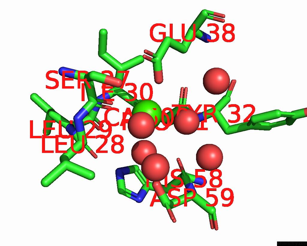
Mono view
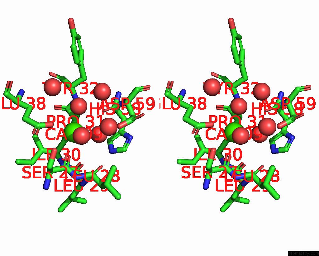
Stereo pair view
|
A full contact list of Calcium with other atoms in the Ca binding
site number 5 of Crystal Structure of Medicago Truncatula Glutamate Dehydrogenase 2 in Complex with 2-Amino-2-Hydroxyglutarate (Reaction Intermediate) and Nad within 5.0Å range:
probe
|
atom
|
residue
|
distance (Å)
|
B
|
Occ
|
E:Ca509
b:18.1
occ:1.00
|
OE2
|
A:GLU38
|
2.2
|
18.7
|
1.0
|
O
|
E:SER27
|
2.2
|
15.6
|
1.0
|
O
|
E:ILE30
|
2.3
|
15.8
|
1.0
|
O
|
E:HOH683
|
2.3
|
20.3
|
1.0
|
O
|
A:HOH868
|
2.4
|
23.8
|
1.0
|
O
|
E:HOH782
|
2.4
|
18.3
|
1.0
|
C
|
E:SER27
|
3.4
|
15.4
|
1.0
|
CD
|
A:GLU38
|
3.4
|
20.1
|
1.0
|
C
|
E:ILE30
|
3.5
|
17.1
|
1.0
|
OE1
|
A:GLU38
|
4.0
|
15.8
|
1.0
|
OG
|
E:SER27
|
4.0
|
18.2
|
1.0
|
CA
|
E:LEU28
|
4.2
|
15.9
|
1.0
|
N
|
E:LEU28
|
4.2
|
15.5
|
1.0
|
N
|
E:ILE30
|
4.2
|
16.7
|
1.0
|
O
|
E:HIS58
|
4.2
|
14.7
|
1.0
|
O
|
E:LEU28
|
4.3
|
19.3
|
1.0
|
CA
|
E:ILE30
|
4.3
|
15.3
|
1.0
|
C
|
E:LEU28
|
4.3
|
17.2
|
1.0
|
N
|
E:TYR32
|
4.3
|
14.7
|
1.0
|
OD2
|
E:ASP59
|
4.3
|
22.2
|
1.0
|
CA
|
E:SER27
|
4.3
|
16.6
|
1.0
|
C
|
E:PRO31
|
4.3
|
14.2
|
1.0
|
CB
|
E:ILE30
|
4.4
|
15.1
|
1.0
|
N
|
E:PRO31
|
4.5
|
14.6
|
1.0
|
CG
|
A:GLU38
|
4.6
|
17.1
|
1.0
|
CA
|
E:PRO31
|
4.6
|
17.0
|
1.0
|
O
|
E:HOH868
|
4.6
|
19.8
|
1.0
|
CB
|
A:GLU38
|
4.7
|
16.4
|
1.0
|
O
|
E:HOH625
|
4.7
|
17.8
|
1.0
|
O
|
A:HOH610
|
4.7
|
27.0
|
1.0
|
CB
|
E:SER27
|
4.8
|
18.4
|
1.0
|
O
|
E:PRO31
|
4.8
|
16.7
|
1.0
|
CA
|
E:TYR32
|
4.9
|
16.0
|
1.0
|
C
|
E:LEU29
|
4.9
|
18.7
|
1.0
|
N
|
E:LEU29
|
4.9
|
17.4
|
1.0
|
CG2
|
E:ILE30
|
5.0
|
19.4
|
1.0
|
|
Calcium binding site 6 out
of 6 in 8s3d
Go back to
Calcium Binding Sites List in 8s3d
Calcium binding site 6 out
of 6 in the Crystal Structure of Medicago Truncatula Glutamate Dehydrogenase 2 in Complex with 2-Amino-2-Hydroxyglutarate (Reaction Intermediate) and Nad
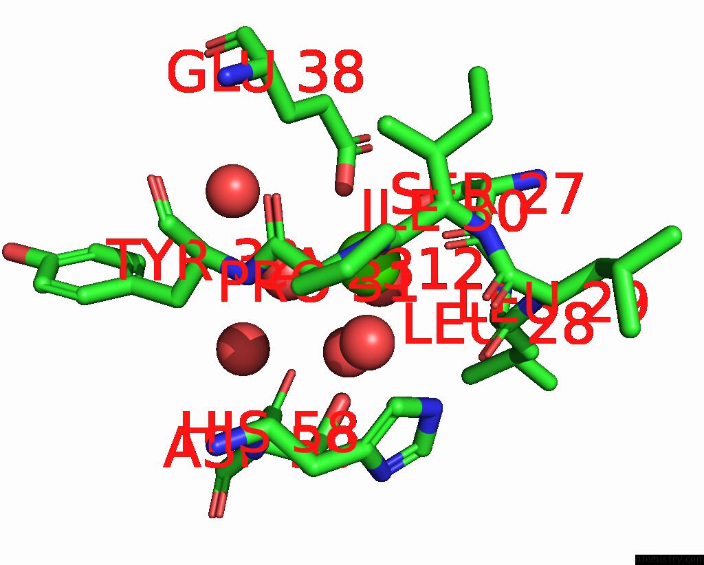
Mono view
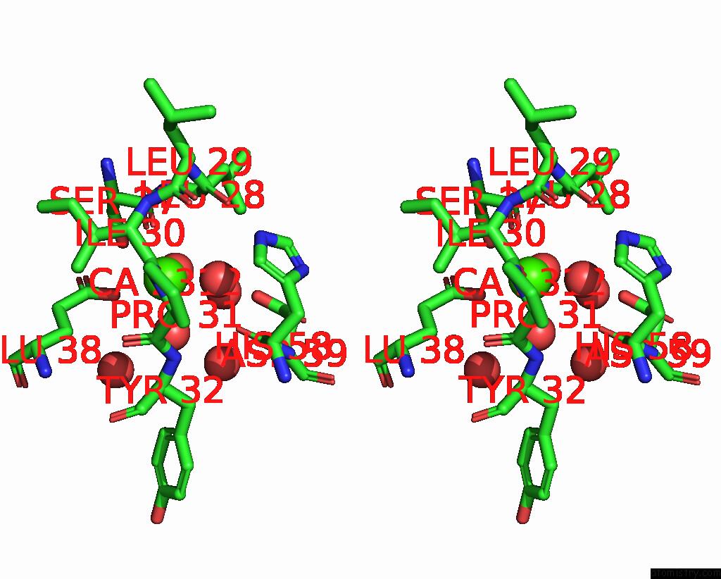
Stereo pair view
|
A full contact list of Calcium with other atoms in the Ca binding
site number 6 of Crystal Structure of Medicago Truncatula Glutamate Dehydrogenase 2 in Complex with 2-Amino-2-Hydroxyglutarate (Reaction Intermediate) and Nad within 5.0Å range:
probe
|
atom
|
residue
|
distance (Å)
|
B
|
Occ
|
F:Ca2312
b:18.0
occ:1.00
|
O
|
F:HOH2498
|
2.2
|
22.5
|
1.0
|
OE2
|
C:GLU38
|
2.3
|
19.4
|
1.0
|
O
|
F:ILE30
|
2.3
|
14.2
|
1.0
|
O
|
C:HOH821
|
2.3
|
23.8
|
1.0
|
O
|
F:SER27
|
2.3
|
16.7
|
1.0
|
O
|
F:HOH2512
|
2.4
|
17.6
|
1.0
|
C
|
F:SER27
|
3.4
|
17.4
|
1.0
|
CD
|
C:GLU38
|
3.5
|
19.8
|
1.0
|
C
|
F:ILE30
|
3.5
|
15.1
|
1.0
|
OG
|
F:SER27
|
4.0
|
16.6
|
1.0
|
OE1
|
C:GLU38
|
4.0
|
19.9
|
1.0
|
CA
|
F:LEU28
|
4.1
|
16.7
|
1.0
|
O
|
F:LEU28
|
4.2
|
17.9
|
1.0
|
N
|
F:LEU28
|
4.2
|
17.0
|
1.0
|
N
|
F:ILE30
|
4.2
|
16.8
|
1.0
|
C
|
F:LEU28
|
4.2
|
15.3
|
1.0
|
CA
|
F:ILE30
|
4.2
|
18.4
|
1.0
|
O
|
F:HIS58
|
4.3
|
15.4
|
1.0
|
N
|
F:TYR32
|
4.3
|
15.0
|
1.0
|
OD2
|
F:ASP59
|
4.3
|
23.5
|
1.0
|
CB
|
F:ILE30
|
4.4
|
16.4
|
1.0
|
C
|
F:PRO31
|
4.4
|
17.3
|
1.0
|
CA
|
F:SER27
|
4.4
|
16.2
|
1.0
|
N
|
F:PRO31
|
4.5
|
16.5
|
1.0
|
CA
|
F:PRO31
|
4.6
|
17.2
|
1.0
|
O
|
C:HOH736
|
4.6
|
24.2
|
1.0
|
O
|
F:HOH2421
|
4.6
|
16.4
|
1.0
|
CG
|
C:GLU38
|
4.6
|
17.1
|
1.0
|
O
|
F:HOH2691
|
4.7
|
19.9
|
1.0
|
CB
|
C:GLU38
|
4.7
|
17.0
|
1.0
|
O
|
F:PRO31
|
4.8
|
17.1
|
1.0
|
CB
|
F:SER27
|
4.8
|
16.5
|
1.0
|
C
|
F:LEU29
|
4.9
|
21.1
|
1.0
|
N
|
F:LEU29
|
4.9
|
17.9
|
1.0
|
CA
|
F:TYR32
|
4.9
|
18.1
|
1.0
|
CG2
|
F:ILE30
|
5.0
|
18.3
|
1.0
|
|
Reference:
M.Grzechowiak,
J.Sliwiak,
A.Link,
M.Ruszkowski.
Legume-Type Glutamate Dehydrogenase: Structure, Activity, and Inhibition Studies. Int.J.Biol.Macromol. V. 278 34648 2024.
ISSN: ISSN 0141-8130
PubMed: 39142482
DOI: 10.1016/J.IJBIOMAC.2024.134648
Page generated: Sat Sep 28 09:03:19 2024
|