Calcium in PDB 8wbr: Crystal Structure of Cis-Epoxysuccinate Hydrolases Klcesh[L]
Protein crystallography data
The structure of Crystal Structure of Cis-Epoxysuccinate Hydrolases Klcesh[L], PDB code: 8wbr
was solved by
S.Dong,
J.S.Xuan,
Y.G.Feng,
Q.Cui,
with X-Ray Crystallography technique. A brief refinement statistics is given in the table below:
| Resolution Low / High (Å) | 30.87 / 2.02 |
| Space group | P 21 21 21 |
| Cell size a, b, c (Å), α, β, γ (°) | 66.3, 84.749, 92.851, 90, 90, 90 |
| R / Rfree (%) | 20.6 / 23.5 |
Calcium Binding Sites:
The binding sites of Calcium atom in the Crystal Structure of Cis-Epoxysuccinate Hydrolases Klcesh[L]
(pdb code 8wbr). This binding sites where shown within
5.0 Angstroms radius around Calcium atom.
In total only one binding site of Calcium was determined in the Crystal Structure of Cis-Epoxysuccinate Hydrolases Klcesh[L], PDB code: 8wbr:
In total only one binding site of Calcium was determined in the Crystal Structure of Cis-Epoxysuccinate Hydrolases Klcesh[L], PDB code: 8wbr:
Calcium binding site 1 out of 1 in 8wbr
Go back to
Calcium binding site 1 out
of 1 in the Crystal Structure of Cis-Epoxysuccinate Hydrolases Klcesh[L]
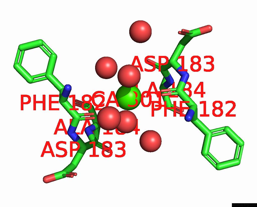
Mono view
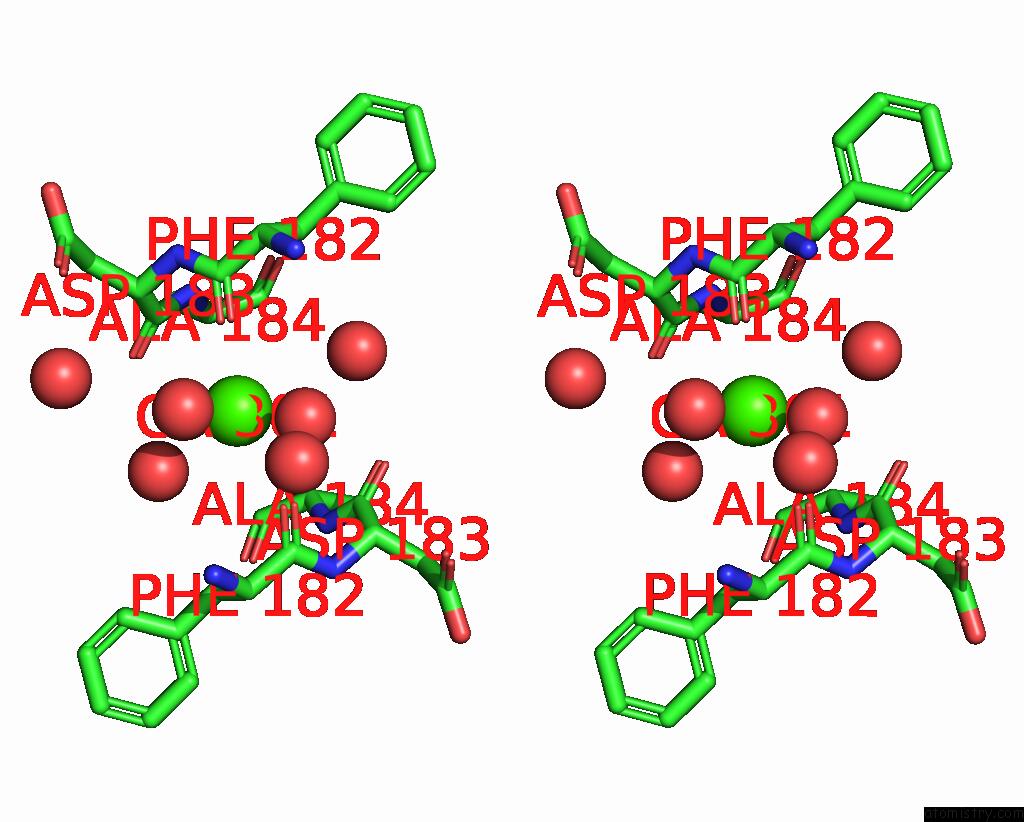
Stereo pair view
Mono view
Stereo pair view
A full contact list of Calcium with other atoms in the Ca binding
site number 1 of Crystal Structure of Cis-Epoxysuccinate Hydrolases Klcesh[L] within 5.0Å range:
|
Reference:
S.Dong,
J.Xuan,
Y.Feng,
Q.Cui.
Deciphering the Stereo-Specific Catalytic Mechanisms of Cis-Epoxysuccinate Hydrolases Producing L(+)-Tartaric Acid. J.Biol.Chem. 05635 2024.
ISSN: ESSN 1083-351X
PubMed: 38199576
DOI: 10.1016/J.JBC.2024.105635
Page generated: Fri Jul 19 12:36:07 2024
ISSN: ESSN 1083-351X
PubMed: 38199576
DOI: 10.1016/J.JBC.2024.105635
Last articles
Zn in 9J0NZn in 9J0O
Zn in 9J0P
Zn in 9FJX
Zn in 9EKB
Zn in 9C0F
Zn in 9CAH
Zn in 9CH0
Zn in 9CH3
Zn in 9CH1