Calcium in PDB 8wg2: Crystal Structure of GH97 Glucodextranase Mutant E509Q From Flavobacterium Johnsoniae in Complex with Isomaltotriose
Enzymatic activity of Crystal Structure of GH97 Glucodextranase Mutant E509Q From Flavobacterium Johnsoniae in Complex with Isomaltotriose
All present enzymatic activity of Crystal Structure of GH97 Glucodextranase Mutant E509Q From Flavobacterium Johnsoniae in Complex with Isomaltotriose:
3.2.1.70;
3.2.1.70;
Protein crystallography data
The structure of Crystal Structure of GH97 Glucodextranase Mutant E509Q From Flavobacterium Johnsoniae in Complex with Isomaltotriose, PDB code: 8wg2
was solved by
R.Kurata,
S.Nakamura,
T.Miyazaki,
with X-Ray Crystallography technique. A brief refinement statistics is given in the table below:
| Resolution Low / High (Å) | 49.16 / 2.45 |
| Space group | P 21 21 2 |
| Cell size a, b, c (Å), α, β, γ (°) | 308.21, 103.62, 95.75, 90, 90, 90 |
| R / Rfree (%) | 18.5 / 23.4 |
Calcium Binding Sites:
The binding sites of Calcium atom in the Crystal Structure of GH97 Glucodextranase Mutant E509Q From Flavobacterium Johnsoniae in Complex with Isomaltotriose
(pdb code 8wg2). This binding sites where shown within
5.0 Angstroms radius around Calcium atom.
In total 4 binding sites of Calcium where determined in the Crystal Structure of GH97 Glucodextranase Mutant E509Q From Flavobacterium Johnsoniae in Complex with Isomaltotriose, PDB code: 8wg2:
Jump to Calcium binding site number: 1; 2; 3; 4;
In total 4 binding sites of Calcium where determined in the Crystal Structure of GH97 Glucodextranase Mutant E509Q From Flavobacterium Johnsoniae in Complex with Isomaltotriose, PDB code: 8wg2:
Jump to Calcium binding site number: 1; 2; 3; 4;
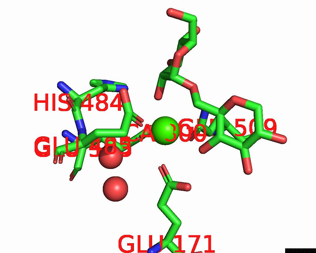
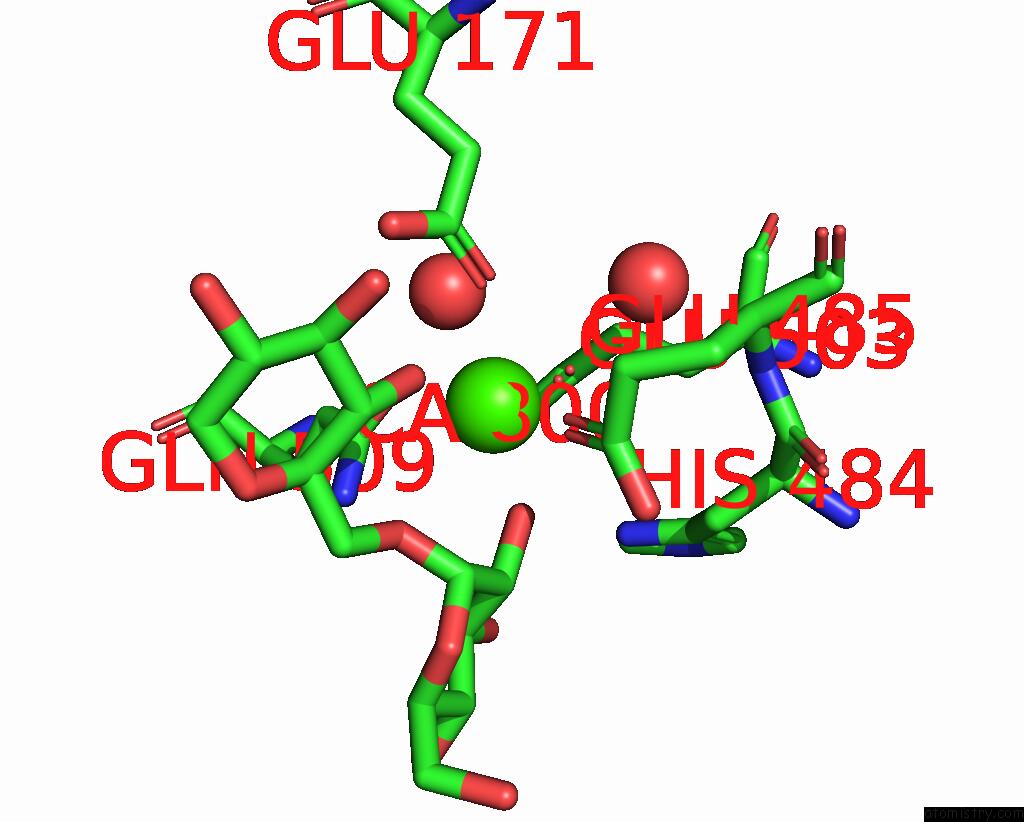
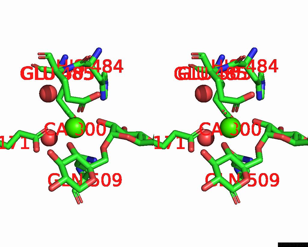
Calcium binding site 1 out of 4 in 8wg2
Go back to
Calcium binding site 1 out
of 4 in the Crystal Structure of GH97 Glucodextranase Mutant E509Q From Flavobacterium Johnsoniae in Complex with Isomaltotriose
Mono view
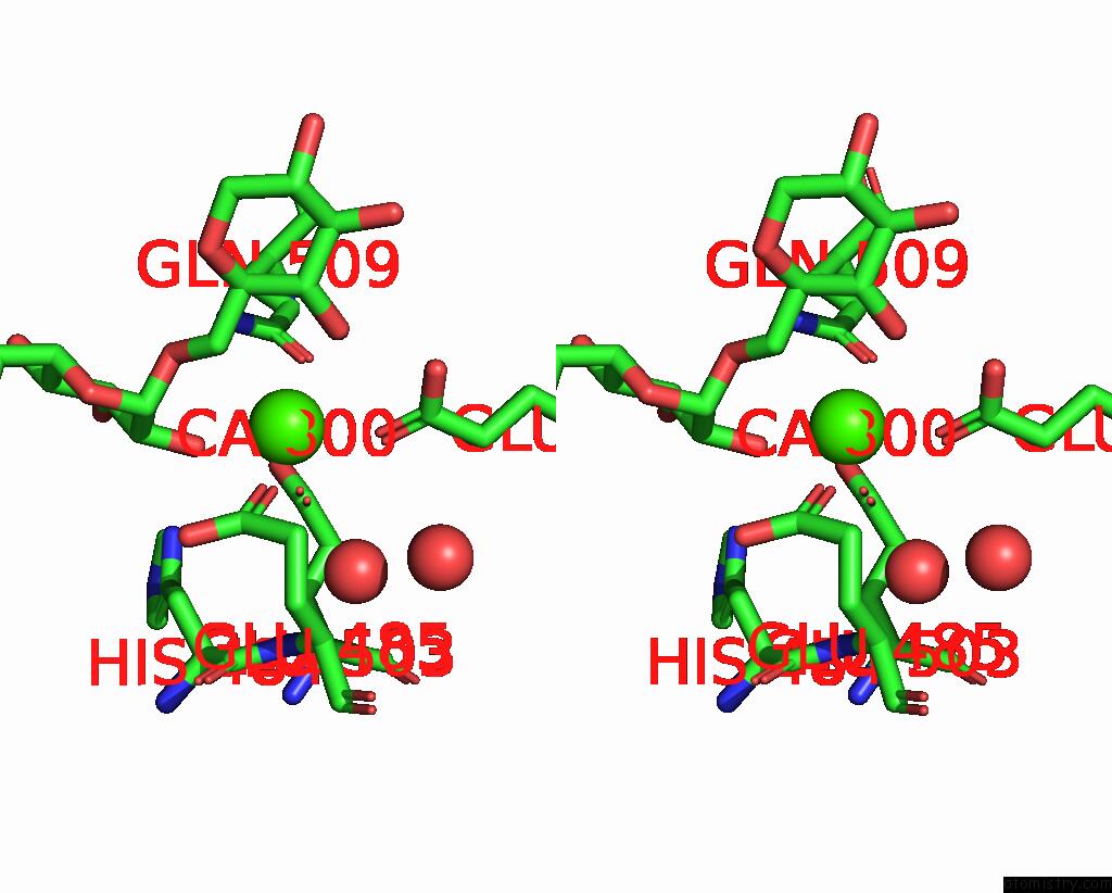
Stereo pair view
Mono view
Stereo pair view
A full contact list of Calcium with other atoms in the Ca binding
site number 1 of Crystal Structure of GH97 Glucodextranase Mutant E509Q From Flavobacterium Johnsoniae in Complex with Isomaltotriose within 5.0Å range:
|
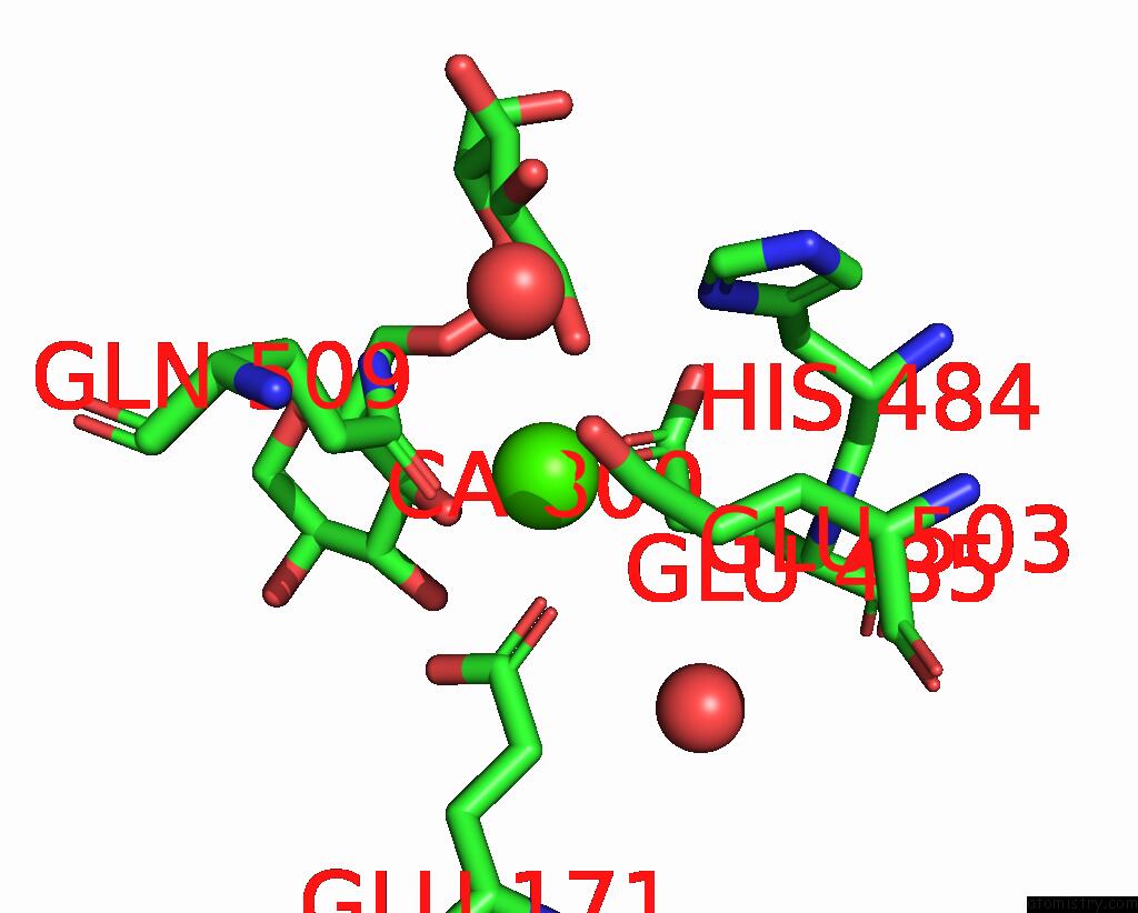
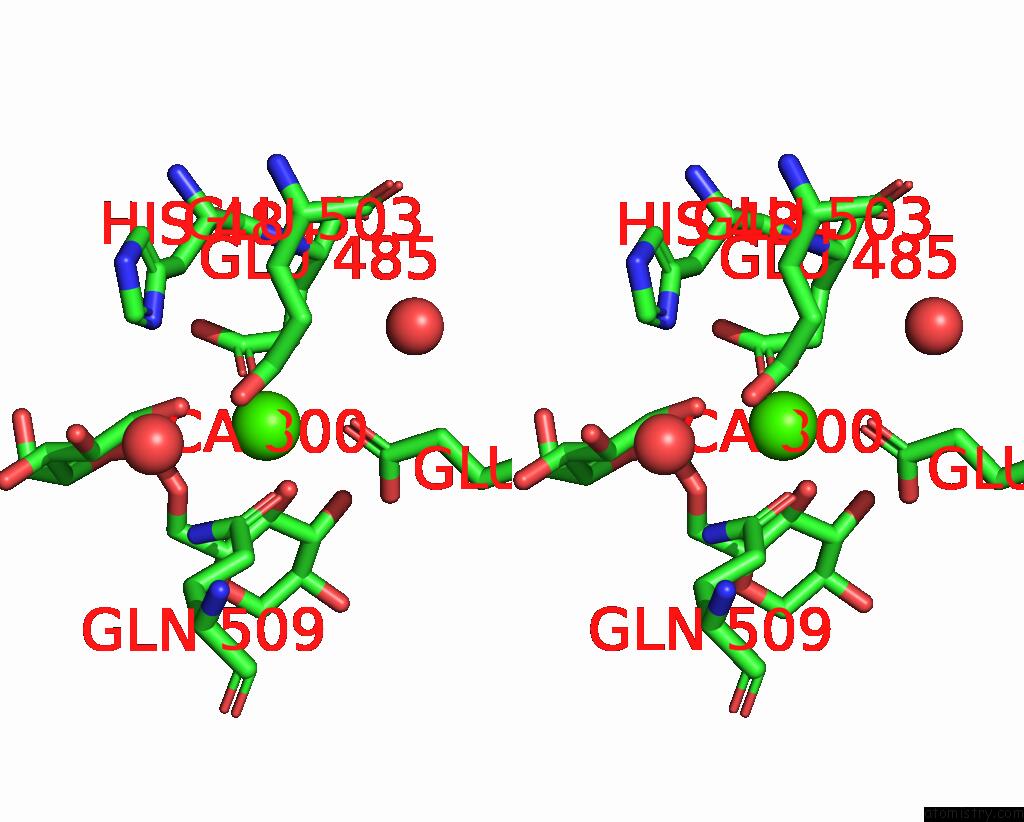
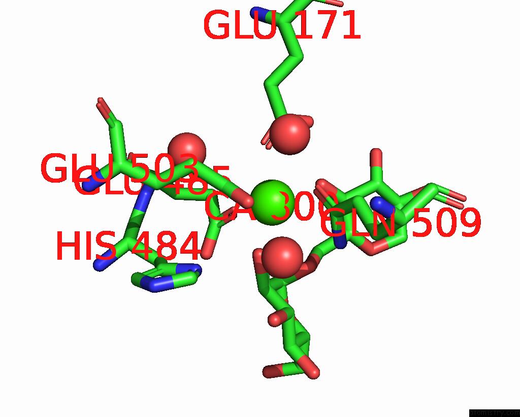
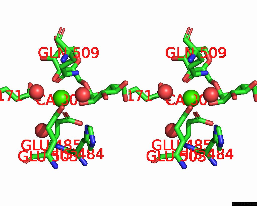
Calcium binding site 2 out of 4 in 8wg2
Go back to
Calcium binding site 2 out
of 4 in the Crystal Structure of GH97 Glucodextranase Mutant E509Q From Flavobacterium Johnsoniae in Complex with Isomaltotriose
Mono view
Stereo pair view
Mono view
Stereo pair view
A full contact list of Calcium with other atoms in the Ca binding
site number 2 of Crystal Structure of GH97 Glucodextranase Mutant E509Q From Flavobacterium Johnsoniae in Complex with Isomaltotriose within 5.0Å range:
|
Calcium binding site 3 out of 4 in 8wg2
Go back to
Calcium binding site 3 out
of 4 in the Crystal Structure of GH97 Glucodextranase Mutant E509Q From Flavobacterium Johnsoniae in Complex with Isomaltotriose
Mono view
Stereo pair view
Mono view
Stereo pair view
A full contact list of Calcium with other atoms in the Ca binding
site number 3 of Crystal Structure of GH97 Glucodextranase Mutant E509Q From Flavobacterium Johnsoniae in Complex with Isomaltotriose within 5.0Å range:
|
Calcium binding site 4 out of 4 in 8wg2
Go back to
Calcium binding site 4 out
of 4 in the Crystal Structure of GH97 Glucodextranase Mutant E509Q From Flavobacterium Johnsoniae in Complex with Isomaltotriose
Mono view
Stereo pair view
Mono view
Stereo pair view
A full contact list of Calcium with other atoms in the Ca binding
site number 4 of Crystal Structure of GH97 Glucodextranase Mutant E509Q From Flavobacterium Johnsoniae in Complex with Isomaltotriose within 5.0Å range:
|
Reference:
S.Nakamura,
R.Kurata,
T.Miyazaki.
Structural Insights Into Alpha-(1->6)-Linkage Preference of GH97 Glucodextranase From Flavobacterium Johnsoniae. Febs J. 2024.
ISSN: ISSN 1742-464X
PubMed: 38661728
DOI: 10.1111/FEBS.17139
Page generated: Fri Jul 19 12:39:21 2024
ISSN: ISSN 1742-464X
PubMed: 38661728
DOI: 10.1111/FEBS.17139
Last articles
Zn in 9J0NZn in 9J0O
Zn in 9J0P
Zn in 9FJX
Zn in 9EKB
Zn in 9C0F
Zn in 9CAH
Zn in 9CH0
Zn in 9CH3
Zn in 9CH1