Calcium »
PDB 1aui-1b82 »
1avr »
Calcium in PDB 1avr: Crystal and Molecular Structure of Human Annexin V After Refinement. Implications For Structure, Membrane Binding and Ion Channel Formation of the Annexin Family of Proteins
Protein crystallography data
The structure of Crystal and Molecular Structure of Human Annexin V After Refinement. Implications For Structure, Membrane Binding and Ion Channel Formation of the Annexin Family of Proteins, PDB code: 1avr
was solved by
R.Huber,
R.Berendes,
A.Burger,
M.Schneider,
A.Karshikov,
H.Luecke,
J.Roemisch,
E.Paques,
with X-Ray Crystallography technique. A brief refinement statistics is given in the table below:
| Resolution Low / High (Å) | N/A / 2.30 |
| Space group | H 3 |
| Cell size a, b, c (Å), α, β, γ (°) | 99.700, 99.700, 96.350, 90.00, 90.00, 120.00 |
| R / Rfree (%) | 18.4 / n/a |
Calcium Binding Sites:
The binding sites of Calcium atom in the Crystal and Molecular Structure of Human Annexin V After Refinement. Implications For Structure, Membrane Binding and Ion Channel Formation of the Annexin Family of Proteins
(pdb code 1avr). This binding sites where shown within
5.0 Angstroms radius around Calcium atom.
In total 5 binding sites of Calcium where determined in the Crystal and Molecular Structure of Human Annexin V After Refinement. Implications For Structure, Membrane Binding and Ion Channel Formation of the Annexin Family of Proteins, PDB code: 1avr:
Jump to Calcium binding site number: 1; 2; 3; 4; 5;
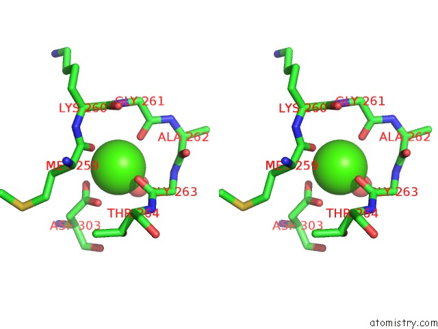
Calcium binding site 1 out of 5 in 1avr
Go back to
Calcium binding site 1 out
of 5 in the Crystal and Molecular Structure of Human Annexin V After Refinement. Implications For Structure, Membrane Binding and Ion Channel Formation of the Annexin Family of Proteins

Mono view

Stereo pair view
Mono view
Stereo pair view
A full contact list of Calcium with other atoms in the Ca binding
site number 1 of Crystal and Molecular Structure of Human Annexin V After Refinement. Implications For Structure, Membrane Binding and Ion Channel Formation of the Annexin Family of Proteins within 5.0Å range:
|
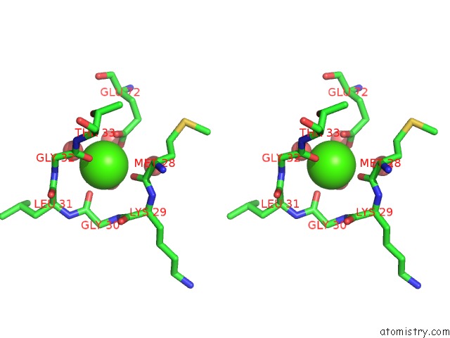
Calcium binding site 2 out of 5 in 1avr
Go back to
Calcium binding site 2 out
of 5 in the Crystal and Molecular Structure of Human Annexin V After Refinement. Implications For Structure, Membrane Binding and Ion Channel Formation of the Annexin Family of Proteins

Mono view
Stereo pair view
Mono view
Stereo pair view
A full contact list of Calcium with other atoms in the Ca binding
site number 2 of Crystal and Molecular Structure of Human Annexin V After Refinement. Implications For Structure, Membrane Binding and Ion Channel Formation of the Annexin Family of Proteins within 5.0Å range:
|
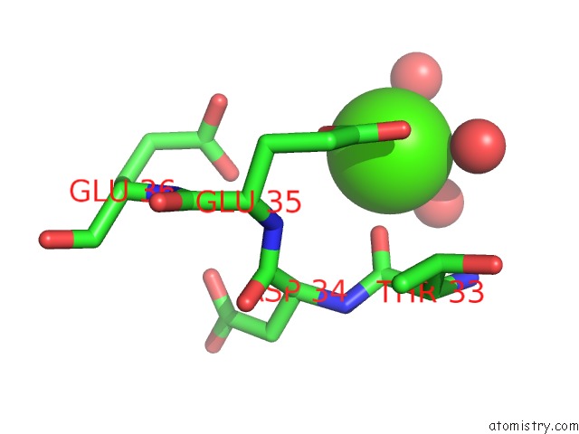
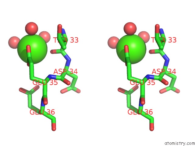
Calcium binding site 3 out of 5 in 1avr
Go back to
Calcium binding site 3 out
of 5 in the Crystal and Molecular Structure of Human Annexin V After Refinement. Implications For Structure, Membrane Binding and Ion Channel Formation of the Annexin Family of Proteins

Mono view
Stereo pair view
Mono view
Stereo pair view
A full contact list of Calcium with other atoms in the Ca binding
site number 3 of Crystal and Molecular Structure of Human Annexin V After Refinement. Implications For Structure, Membrane Binding and Ion Channel Formation of the Annexin Family of Proteins within 5.0Å range:
|
Calcium binding site 4 out of 5 in 1avr
Go back to
Calcium binding site 4 out
of 5 in the Crystal and Molecular Structure of Human Annexin V After Refinement. Implications For Structure, Membrane Binding and Ion Channel Formation of the Annexin Family of Proteins
Mono view
Stereo pair view
Mono view
Stereo pair view
A full contact list of Calcium with other atoms in the Ca binding
site number 4 of Crystal and Molecular Structure of Human Annexin V After Refinement. Implications For Structure, Membrane Binding and Ion Channel Formation of the Annexin Family of Proteins within 5.0Å range:
|
Calcium binding site 5 out of 5 in 1avr
Go back to
Calcium binding site 5 out
of 5 in the Crystal and Molecular Structure of Human Annexin V After Refinement. Implications For Structure, Membrane Binding and Ion Channel Formation of the Annexin Family of Proteins

Mono view

Stereo pair view
Mono view
Stereo pair view
A full contact list of Calcium with other atoms in the Ca binding
site number 5 of Crystal and Molecular Structure of Human Annexin V After Refinement. Implications For Structure, Membrane Binding and Ion Channel Formation of the Annexin Family of Proteins within 5.0Å range:
|
Reference:
R.Huber,
R.Berendes,
A.Burger,
M.Schneider,
A.Karshikov,
H.Luecke,
J.Romisch,
E.Paques.
Crystal and Molecular Structure of Human Annexin V After Refinement. Implications For Structure, Membrane Binding and Ion Channel Formation of the Annexin Family of Proteins. J.Mol.Biol. V. 223 683 1992.
ISSN: ISSN 0022-2836
PubMed: 1311770
DOI: 10.1016/0022-2836(92)90984-R
Page generated: Mon Jul 7 13:29:22 2025
ISSN: ISSN 0022-2836
PubMed: 1311770
DOI: 10.1016/0022-2836(92)90984-R
Last articles
Cl in 8B78Cl in 8B6T
Cl in 8B6S
Cl in 8B75
Cl in 8B6P
Cl in 8B6R
Cl in 8B6Q
Cl in 8B6I
Cl in 8B6O
Cl in 8B5X