Calcium »
PDB 1aui-1b82 »
1b0p »
Calcium in PDB 1b0p: Crystal Structure of Pyruvate-Ferredoxin Oxidoreductase From Desulfovibrio Africanus
Enzymatic activity of Crystal Structure of Pyruvate-Ferredoxin Oxidoreductase From Desulfovibrio Africanus
All present enzymatic activity of Crystal Structure of Pyruvate-Ferredoxin Oxidoreductase From Desulfovibrio Africanus:
1.2.7.1;
1.2.7.1;
Protein crystallography data
The structure of Crystal Structure of Pyruvate-Ferredoxin Oxidoreductase From Desulfovibrio Africanus, PDB code: 1b0p
was solved by
E.Chabriere,
M.H.Charon,
A.Volbeda,
with X-Ray Crystallography technique. A brief refinement statistics is given in the table below:
| Resolution Low / High (Å) | 6.00 / 2.31 |
| Space group | P 21 21 21 |
| Cell size a, b, c (Å), α, β, γ (°) | 84.800, 144.900, 203.000, 90.00, 90.00, 90.00 |
| R / Rfree (%) | 19.9 / 27.1 |
Other elements in 1b0p:
The structure of Crystal Structure of Pyruvate-Ferredoxin Oxidoreductase From Desulfovibrio Africanus also contains other interesting chemical elements:
| Magnesium | (Mg) | 2 atoms |
| Iron | (Fe) | 24 atoms |
Calcium Binding Sites:
The binding sites of Calcium atom in the Crystal Structure of Pyruvate-Ferredoxin Oxidoreductase From Desulfovibrio Africanus
(pdb code 1b0p). This binding sites where shown within
5.0 Angstroms radius around Calcium atom.
In total 2 binding sites of Calcium where determined in the Crystal Structure of Pyruvate-Ferredoxin Oxidoreductase From Desulfovibrio Africanus, PDB code: 1b0p:
Jump to Calcium binding site number: 1; 2;
In total 2 binding sites of Calcium where determined in the Crystal Structure of Pyruvate-Ferredoxin Oxidoreductase From Desulfovibrio Africanus, PDB code: 1b0p:
Jump to Calcium binding site number: 1; 2;
Calcium binding site 1 out of 2 in 1b0p
Go back to
Calcium binding site 1 out
of 2 in the Crystal Structure of Pyruvate-Ferredoxin Oxidoreductase From Desulfovibrio Africanus
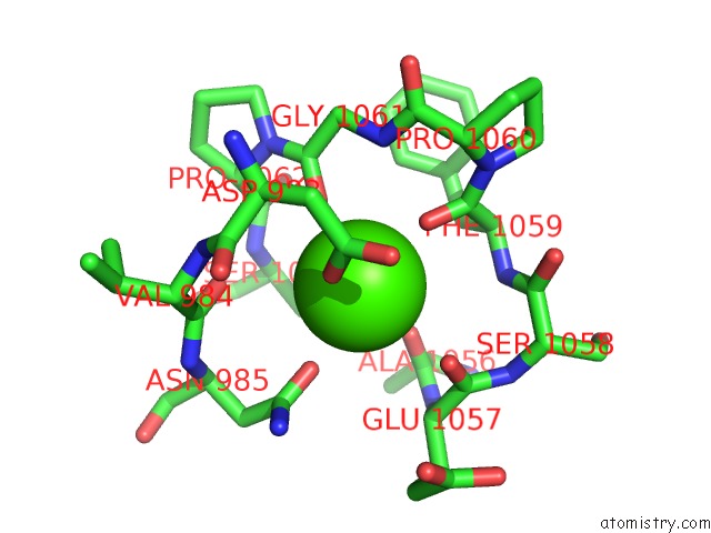
Mono view
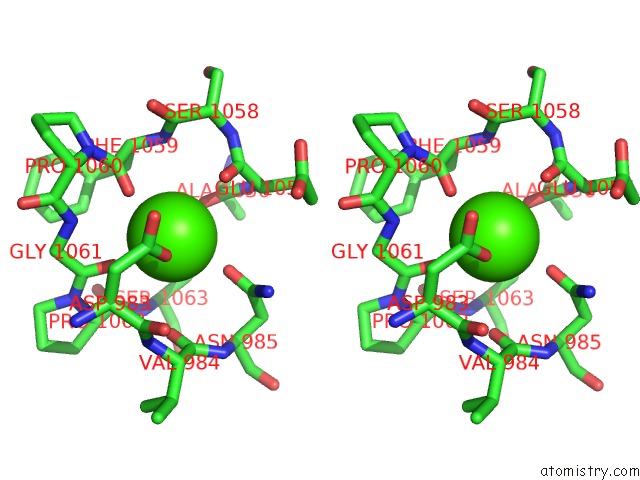
Stereo pair view
Mono view
Stereo pair view
A full contact list of Calcium with other atoms in the Ca binding
site number 1 of Crystal Structure of Pyruvate-Ferredoxin Oxidoreductase From Desulfovibrio Africanus within 5.0Å range:
|
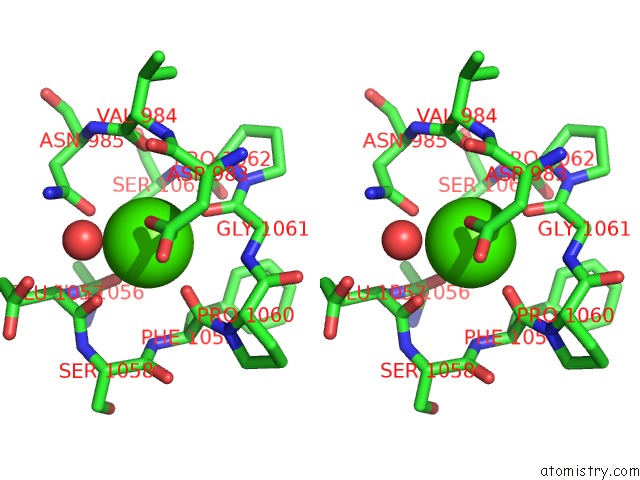
Calcium binding site 2 out of 2 in 1b0p
Go back to
Calcium binding site 2 out
of 2 in the Crystal Structure of Pyruvate-Ferredoxin Oxidoreductase From Desulfovibrio Africanus
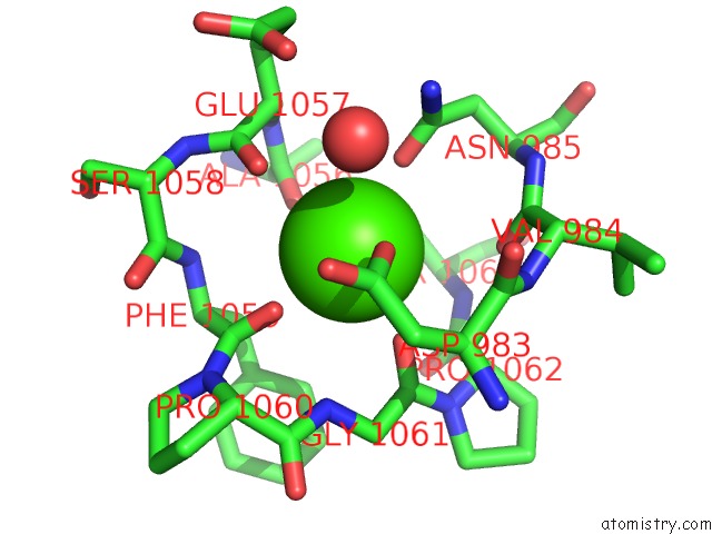
Mono view
Stereo pair view
Mono view
Stereo pair view
A full contact list of Calcium with other atoms in the Ca binding
site number 2 of Crystal Structure of Pyruvate-Ferredoxin Oxidoreductase From Desulfovibrio Africanus within 5.0Å range:
|
Reference:
E.Chabriere,
M.H.Charon,
A.Volbeda,
L.Pieulle,
E.C.Hatchikian,
J.C.Fontecilla-Camps.
Crystal Structures of the Key Anaerobic Enzyme Pyruvate:Ferredoxin Oxidoreductase, Free and in Complex with Pyruvate. Nat.Struct.Biol. V. 6 182 1999.
ISSN: ISSN 1072-8368
PubMed: 10048931
DOI: 10.1038/5870
Page generated: Mon Jul 7 13:33:57 2025
ISSN: ISSN 1072-8368
PubMed: 10048931
DOI: 10.1038/5870
Last articles
Cl in 5RKECl in 5RH4
Cl in 5RH3
Cl in 5RH2
Cl in 5RFZ
Cl in 5RH1
Cl in 5RFU
Cl in 5RFP
Cl in 5RFH
Cl in 5RET