Calcium »
PDB 1aui-1b82 »
1b2y »
Calcium in PDB 1b2y: Structure of Human Pancreatic Alpha-Amylase in Complex with the Carbohydrate Inhibitor Acarbose
Enzymatic activity of Structure of Human Pancreatic Alpha-Amylase in Complex with the Carbohydrate Inhibitor Acarbose
All present enzymatic activity of Structure of Human Pancreatic Alpha-Amylase in Complex with the Carbohydrate Inhibitor Acarbose:
3.2.1.1;
3.2.1.1;
Protein crystallography data
The structure of Structure of Human Pancreatic Alpha-Amylase in Complex with the Carbohydrate Inhibitor Acarbose, PDB code: 1b2y
was solved by
V.Nahoum,
F.Payan,
with X-Ray Crystallography technique. A brief refinement statistics is given in the table below:
| Resolution Low / High (Å) | 11.00 / 3.20 |
| Space group | P 21 21 21 |
| Cell size a, b, c (Å), α, β, γ (°) | 53.110, 75.100, 137.130, 90.00, 90.00, 90.00 |
| R / Rfree (%) | 19.1 / 21.7 |
Other elements in 1b2y:
The structure of Structure of Human Pancreatic Alpha-Amylase in Complex with the Carbohydrate Inhibitor Acarbose also contains other interesting chemical elements:
| Chlorine | (Cl) | 1 atom |
Calcium Binding Sites:
The binding sites of Calcium atom in the Structure of Human Pancreatic Alpha-Amylase in Complex with the Carbohydrate Inhibitor Acarbose
(pdb code 1b2y). This binding sites where shown within
5.0 Angstroms radius around Calcium atom.
In total only one binding site of Calcium was determined in the Structure of Human Pancreatic Alpha-Amylase in Complex with the Carbohydrate Inhibitor Acarbose, PDB code: 1b2y:
In total only one binding site of Calcium was determined in the Structure of Human Pancreatic Alpha-Amylase in Complex with the Carbohydrate Inhibitor Acarbose, PDB code: 1b2y:
Calcium binding site 1 out of 1 in 1b2y
Go back to
Calcium binding site 1 out
of 1 in the Structure of Human Pancreatic Alpha-Amylase in Complex with the Carbohydrate Inhibitor Acarbose
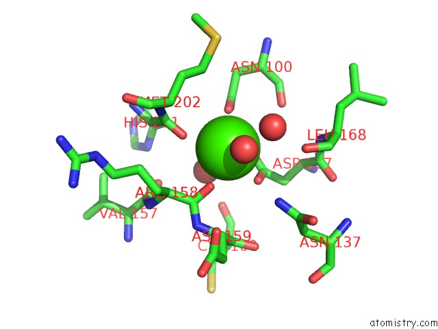
Mono view
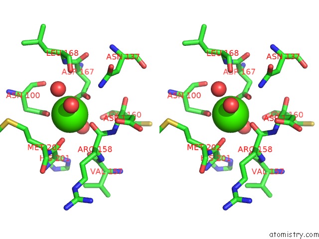
Stereo pair view
Mono view
Stereo pair view
A full contact list of Calcium with other atoms in the Ca binding
site number 1 of Structure of Human Pancreatic Alpha-Amylase in Complex with the Carbohydrate Inhibitor Acarbose within 5.0Å range:
|
Reference:
V.Nahoum,
G.Roux,
V.Anton,
P.Rouge,
A.Puigserver,
H.Bischoff,
B.Henrissat,
F.Payan.
Crystal Structures of Human Pancreatic Alpha-Amylase in Complex with Carbohydrate and Proteinaceous Inhibitors. Biochem.J. V.Pt 1 201 2000.
ISSN: ISSN 0264-6021
PubMed: 10657258
DOI: 10.1042/0264-6021:3460201
Page generated: Thu Jul 11 06:14:12 2024
ISSN: ISSN 0264-6021
PubMed: 10657258
DOI: 10.1042/0264-6021:3460201
Last articles
Zn in 9J0NZn in 9J0O
Zn in 9J0P
Zn in 9FJX
Zn in 9EKB
Zn in 9C0F
Zn in 9CAH
Zn in 9CH0
Zn in 9CH3
Zn in 9CH1