Calcium »
PDB 1aui-1b82 »
1b47 »
Calcium in PDB 1b47: Structure of the N-Terminal Domain of Cbl in Complex with Its Binding Site in Zap-70
Protein crystallography data
The structure of Structure of the N-Terminal Domain of Cbl in Complex with Its Binding Site in Zap-70, PDB code: 1b47
was solved by
W.Meng,
S.Sawasdikosol,
S.J.Burakoff,
M.J.Eck,
with X-Ray Crystallography technique. A brief refinement statistics is given in the table below:
| Resolution Low / High (Å) | 50.00 / 2.20 |
| Space group | C 1 2 1 |
| Cell size a, b, c (Å), α, β, γ (°) | 159.960, 105.480, 84.920, 90.00, 92.06, 90.00 |
| R / Rfree (%) | 21.8 / 26.6 |
Calcium Binding Sites:
The binding sites of Calcium atom in the Structure of the N-Terminal Domain of Cbl in Complex with Its Binding Site in Zap-70
(pdb code 1b47). This binding sites where shown within
5.0 Angstroms radius around Calcium atom.
In total 3 binding sites of Calcium where determined in the Structure of the N-Terminal Domain of Cbl in Complex with Its Binding Site in Zap-70, PDB code: 1b47:
Jump to Calcium binding site number: 1; 2; 3;
In total 3 binding sites of Calcium where determined in the Structure of the N-Terminal Domain of Cbl in Complex with Its Binding Site in Zap-70, PDB code: 1b47:
Jump to Calcium binding site number: 1; 2; 3;
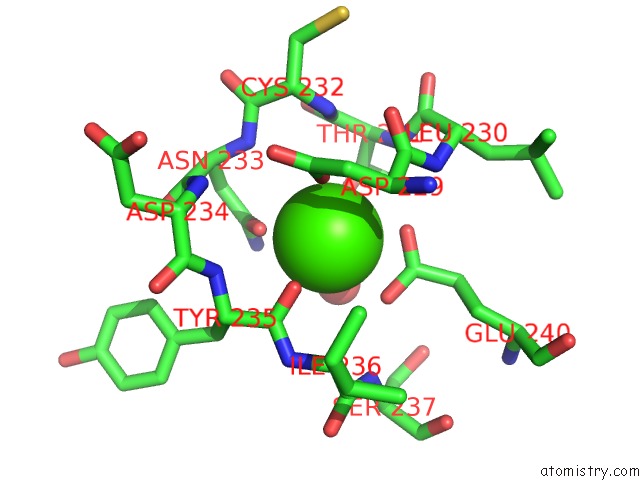
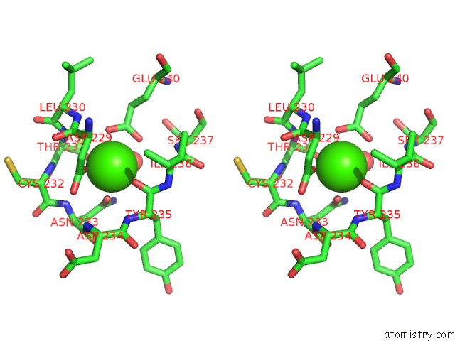
Calcium binding site 1 out of 3 in 1b47
Go back to
Calcium binding site 1 out
of 3 in the Structure of the N-Terminal Domain of Cbl in Complex with Its Binding Site in Zap-70
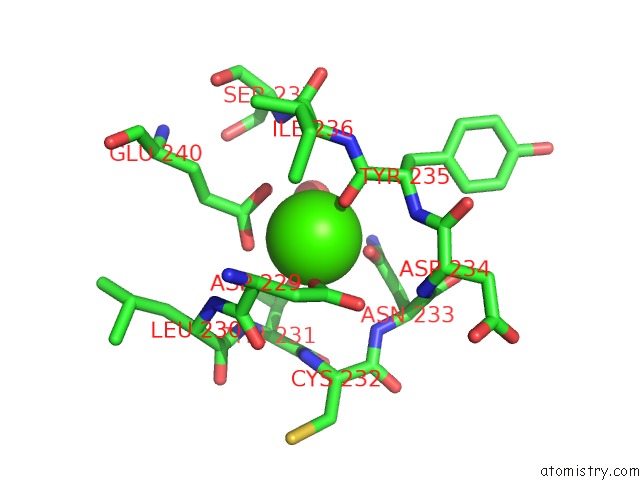
Mono view
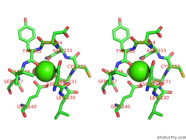
Stereo pair view
Mono view
Stereo pair view
A full contact list of Calcium with other atoms in the Ca binding
site number 1 of Structure of the N-Terminal Domain of Cbl in Complex with Its Binding Site in Zap-70 within 5.0Å range:
|
Calcium binding site 2 out of 3 in 1b47
Go back to
Calcium binding site 2 out
of 3 in the Structure of the N-Terminal Domain of Cbl in Complex with Its Binding Site in Zap-70
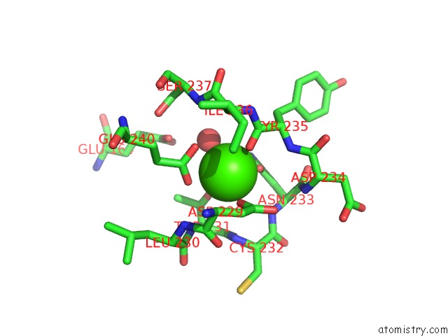
Mono view
Stereo pair view
Mono view
Stereo pair view
A full contact list of Calcium with other atoms in the Ca binding
site number 2 of Structure of the N-Terminal Domain of Cbl in Complex with Its Binding Site in Zap-70 within 5.0Å range:
|
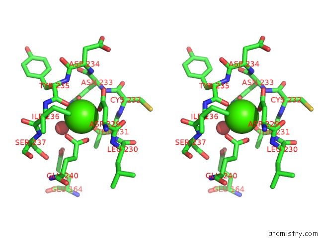
Calcium binding site 3 out of 3 in 1b47
Go back to
Calcium binding site 3 out
of 3 in the Structure of the N-Terminal Domain of Cbl in Complex with Its Binding Site in Zap-70
Mono view
Stereo pair view
Mono view
Stereo pair view
A full contact list of Calcium with other atoms in the Ca binding
site number 3 of Structure of the N-Terminal Domain of Cbl in Complex with Its Binding Site in Zap-70 within 5.0Å range:
|
Reference:
W.Meng,
S.Sawasdikosol,
S.J.Burakoff,
M.J.Eck.
Structure of the Amino-Terminal Domain of Cbl Complexed to Its Binding Site on Zap-70 Kinase. Nature V. 398 84 1999.
ISSN: ISSN 0028-0836
PubMed: 10078535
DOI: 10.1038/18050
Page generated: Mon Jul 7 13:35:04 2025
ISSN: ISSN 0028-0836
PubMed: 10078535
DOI: 10.1038/18050
Last articles
Cl in 5HL3Cl in 5HK9
Cl in 5HKY
Cl in 5HKG
Cl in 5HK7
Cl in 5HJS
Cl in 5HJY
Cl in 5HJP
Cl in 5HJ2
Cl in 5HI6