Calcium in PDB 1bch: Mannose-Binding Protein-A Mutant (Qpdwgh) Complexed with N- Acetyl-D-Galactosamine
Protein crystallography data
The structure of Mannose-Binding Protein-A Mutant (Qpdwgh) Complexed with N- Acetyl-D-Galactosamine, PDB code: 1bch
was solved by
A.R.Kolatkar,
W.I.Weis,
with X-Ray Crystallography technique. A brief refinement statistics is given in the table below:
Resolution Low / High (Å)
|
30.00 /
2.00
|
Space group
|
C 1 2 1
|
Cell size a, b, c (Å), α, β, γ (°)
|
80.490,
85.010,
98.710,
90.00,
104.82,
90.00
|
R / Rfree (%)
|
21.9 /
25.2
|
Other elements in 1bch:
The structure of Mannose-Binding Protein-A Mutant (Qpdwgh) Complexed with N- Acetyl-D-Galactosamine also contains other interesting chemical elements:
Calcium Binding Sites:
The binding sites of Calcium atom in the Mannose-Binding Protein-A Mutant (Qpdwgh) Complexed with N- Acetyl-D-Galactosamine
(pdb code 1bch). This binding sites where shown within
5.0 Angstroms radius around Calcium atom.
In total 9 binding sites of Calcium where determined in the
Mannose-Binding Protein-A Mutant (Qpdwgh) Complexed with N- Acetyl-D-Galactosamine, PDB code: 1bch:
Jump to Calcium binding site number:
1;
2;
3;
4;
5;
6;
7;
8;
9;
Calcium binding site 1 out
of 9 in 1bch
Go back to
Calcium Binding Sites List in 1bch
Calcium binding site 1 out
of 9 in the Mannose-Binding Protein-A Mutant (Qpdwgh) Complexed with N- Acetyl-D-Galactosamine
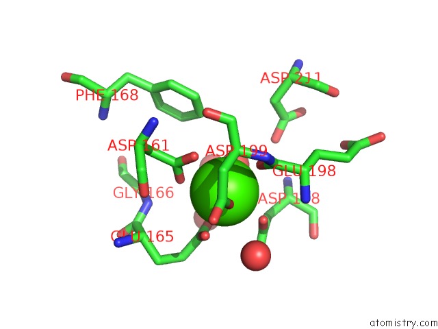
Mono view
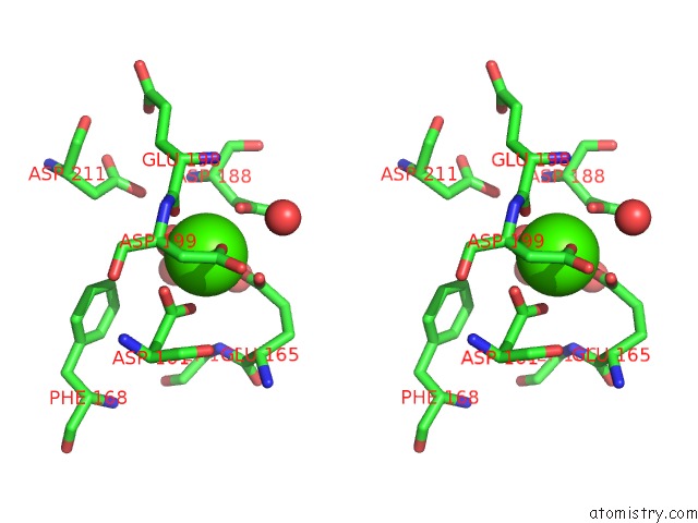
Stereo pair view
|
A full contact list of Calcium with other atoms in the Ca binding
site number 1 of Mannose-Binding Protein-A Mutant (Qpdwgh) Complexed with N- Acetyl-D-Galactosamine within 5.0Å range:
probe
|
atom
|
residue
|
distance (Å)
|
B
|
Occ
|
1:Ca227
b:26.5
occ:1.00
|
O
|
1:HOH241
|
2.4
|
22.8
|
1.0
|
O
|
1:GLU198
|
2.4
|
22.2
|
1.0
|
OD1
|
1:ASP199
|
2.4
|
22.0
|
1.0
|
OD2
|
1:ASP161
|
2.4
|
23.6
|
1.0
|
OE1
|
1:GLU165
|
2.5
|
27.8
|
1.0
|
OD1
|
1:ASP188
|
2.5
|
27.8
|
1.0
|
OE2
|
1:GLU165
|
2.6
|
27.3
|
1.0
|
OD1
|
1:ASP161
|
2.7
|
22.0
|
1.0
|
CD
|
1:GLU165
|
2.9
|
27.2
|
1.0
|
CG
|
1:ASP161
|
2.9
|
24.7
|
1.0
|
CG
|
1:ASP188
|
3.3
|
29.7
|
1.0
|
C
|
1:GLU198
|
3.4
|
23.8
|
1.0
|
CG
|
1:ASP199
|
3.6
|
21.8
|
1.0
|
CA
|
1:ASP199
|
3.8
|
22.5
|
1.0
|
CB
|
1:ASP188
|
3.8
|
29.4
|
1.0
|
N
|
1:ASP199
|
3.9
|
21.2
|
1.0
|
O
|
1:HOH274
|
4.0
|
35.5
|
1.0
|
OD2
|
1:ASP188
|
4.1
|
27.6
|
1.0
|
CB
|
1:ASP199
|
4.2
|
21.8
|
1.0
|
N
|
1:GLU198
|
4.3
|
27.1
|
1.0
|
CA
|
1:ASP188
|
4.4
|
31.2
|
1.0
|
CG
|
1:GLU165
|
4.4
|
26.7
|
1.0
|
CB
|
1:ASP161
|
4.4
|
26.0
|
1.0
|
CA
|
1:GLU198
|
4.5
|
25.9
|
1.0
|
OD2
|
1:ASP199
|
4.5
|
23.2
|
1.0
|
O
|
1:HOH275
|
4.6
|
31.7
|
1.0
|
CZ
|
1:PHE168
|
4.6
|
26.8
|
1.0
|
N
|
1:GLY166
|
4.6
|
27.1
|
1.0
|
O
|
1:ASP161
|
4.7
|
27.4
|
1.0
|
OD2
|
1:ASP211
|
4.7
|
24.3
|
1.0
|
O
|
1:HOH242
|
4.7
|
26.3
|
1.0
|
CA
|
1:GLU165
|
4.9
|
26.0
|
1.0
|
C
|
1:ASP199
|
5.0
|
22.5
|
1.0
|
|
Calcium binding site 2 out
of 9 in 1bch
Go back to
Calcium Binding Sites List in 1bch
Calcium binding site 2 out
of 9 in the Mannose-Binding Protein-A Mutant (Qpdwgh) Complexed with N- Acetyl-D-Galactosamine
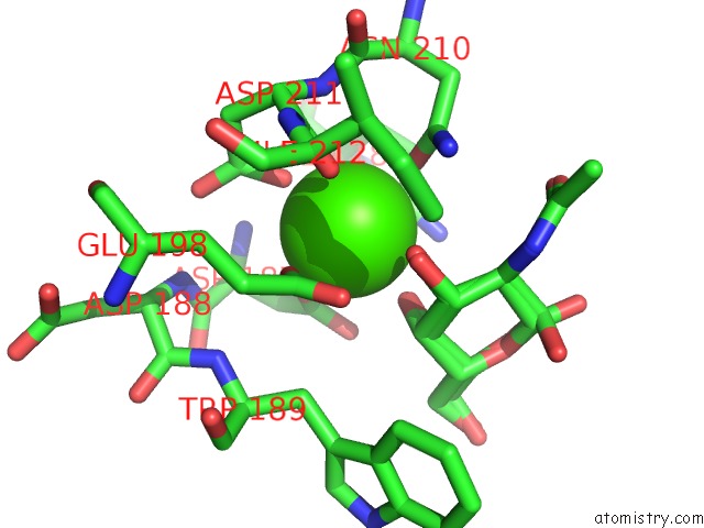
Mono view
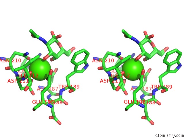
Stereo pair view
|
A full contact list of Calcium with other atoms in the Ca binding
site number 2 of Mannose-Binding Protein-A Mutant (Qpdwgh) Complexed with N- Acetyl-D-Galactosamine within 5.0Å range:
probe
|
atom
|
residue
|
distance (Å)
|
B
|
Occ
|
1:Ca2
b:28.9
occ:1.00
|
OD1
|
1:ASP211
|
2.3
|
24.1
|
1.0
|
OE1
|
1:GLU198
|
2.4
|
26.4
|
1.0
|
OD1
|
1:ASP187
|
2.5
|
28.8
|
1.0
|
O3
|
1:A2G1
|
2.5
|
25.3
|
0.3
|
O
|
1:ASP211
|
2.5
|
24.6
|
1.0
|
OD1
|
1:ASN210
|
2.5
|
25.1
|
1.0
|
OE1
|
1:GLN185
|
2.6
|
28.8
|
1.0
|
O4
|
1:A2G1
|
2.6
|
23.5
|
0.3
|
O4
|
1:A2G1
|
2.6
|
20.1
|
0.3
|
O3
|
1:A2G1
|
2.6
|
22.5
|
0.3
|
CD
|
1:GLU198
|
3.3
|
24.9
|
1.0
|
C4
|
1:A2G1
|
3.3
|
24.5
|
0.3
|
CG
|
1:ASP211
|
3.3
|
26.2
|
1.0
|
CD
|
1:GLN185
|
3.4
|
30.0
|
1.0
|
C3
|
1:A2G1
|
3.4
|
25.1
|
0.3
|
CG
|
1:ASP187
|
3.4
|
30.0
|
1.0
|
CG
|
1:ASN210
|
3.4
|
25.6
|
1.0
|
C4
|
1:A2G1
|
3.5
|
21.6
|
0.3
|
C3
|
1:A2G1
|
3.6
|
21.8
|
0.3
|
OE2
|
1:GLU198
|
3.6
|
27.6
|
1.0
|
C
|
1:ASP211
|
3.6
|
24.3
|
1.0
|
ND2
|
1:ASN210
|
3.8
|
22.9
|
1.0
|
N
|
1:ASP211
|
3.8
|
25.2
|
1.0
|
OD2
|
1:ASP187
|
3.8
|
27.7
|
1.0
|
NE2
|
1:GLN185
|
3.8
|
29.0
|
1.0
|
OD2
|
1:ASP211
|
4.1
|
24.3
|
1.0
|
CA
|
1:ASP211
|
4.1
|
23.7
|
1.0
|
C2
|
1:A2G1
|
4.2
|
24.6
|
0.3
|
N
|
1:ASP187
|
4.2
|
32.0
|
1.0
|
C2
|
1:A2G1
|
4.3
|
21.2
|
0.3
|
CB
|
1:ASP211
|
4.3
|
24.4
|
1.0
|
CG
|
1:GLN185
|
4.5
|
27.7
|
1.0
|
CB
|
1:TRP189
|
4.6
|
37.6
|
1.0
|
CG
|
1:GLU198
|
4.6
|
25.6
|
1.0
|
C
|
1:ASN210
|
4.6
|
26.3
|
1.0
|
CB
|
1:ASP187
|
4.7
|
31.7
|
1.0
|
N
|
1:ILE212
|
4.7
|
24.9
|
1.0
|
C5
|
1:A2G1
|
4.8
|
26.1
|
0.3
|
CB
|
1:ASN210
|
4.8
|
24.6
|
1.0
|
CB
|
1:GLU198
|
4.8
|
24.7
|
1.0
|
N
|
1:TRP189
|
4.8
|
34.0
|
1.0
|
N
|
1:ASP188
|
4.9
|
29.9
|
1.0
|
C5
|
1:A2G1
|
4.9
|
22.6
|
0.3
|
CA
|
1:ASP187
|
4.9
|
30.8
|
1.0
|
CA
|
1:ASN210
|
5.0
|
26.6
|
1.0
|
|
Calcium binding site 3 out
of 9 in 1bch
Go back to
Calcium Binding Sites List in 1bch
Calcium binding site 3 out
of 9 in the Mannose-Binding Protein-A Mutant (Qpdwgh) Complexed with N- Acetyl-D-Galactosamine
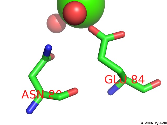
Mono view
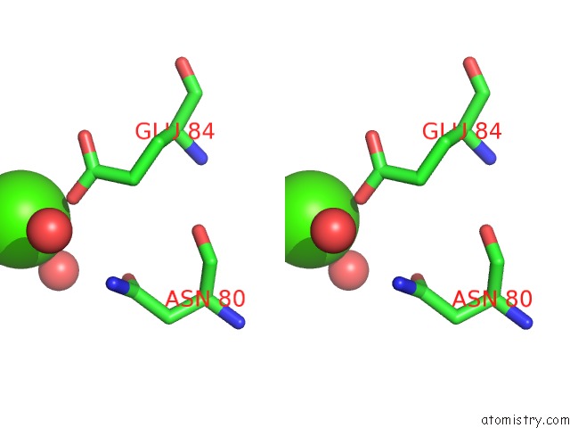
Stereo pair view
|
A full contact list of Calcium with other atoms in the Ca binding
site number 3 of Mannose-Binding Protein-A Mutant (Qpdwgh) Complexed with N- Acetyl-D-Galactosamine within 5.0Å range:
probe
|
atom
|
residue
|
distance (Å)
|
B
|
Occ
|
1:Ca3
b:25.5
occ:1.00
|
OE2
|
1:GLU84
|
2.4
|
25.5
|
1.0
|
O
|
1:HOH228
|
2.5
|
19.8
|
1.0
|
CD
|
1:GLU84
|
3.5
|
26.7
|
1.0
|
OE1
|
1:GLU84
|
4.0
|
27.3
|
1.0
|
OD1
|
1:ASN80
|
4.5
|
23.9
|
1.0
|
ND2
|
1:ASN80
|
4.6
|
23.6
|
1.0
|
O
|
2:HOH234
|
4.6
|
31.2
|
1.0
|
CG
|
1:GLU84
|
4.7
|
26.3
|
1.0
|
CG
|
1:ASN80
|
5.0
|
24.4
|
1.0
|
|
Calcium binding site 4 out
of 9 in 1bch
Go back to
Calcium Binding Sites List in 1bch
Calcium binding site 4 out
of 9 in the Mannose-Binding Protein-A Mutant (Qpdwgh) Complexed with N- Acetyl-D-Galactosamine
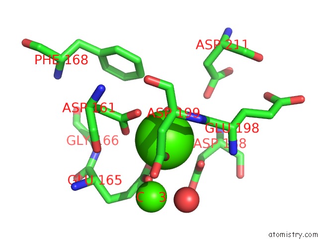
Mono view
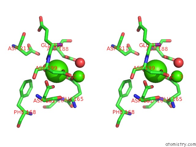
Stereo pair view
|
A full contact list of Calcium with other atoms in the Ca binding
site number 4 of Mannose-Binding Protein-A Mutant (Qpdwgh) Complexed with N- Acetyl-D-Galactosamine within 5.0Å range:
probe
|
atom
|
residue
|
distance (Å)
|
B
|
Occ
|
2:Ca227
b:34.1
occ:1.00
|
O
|
2:HOH272
|
2.3
|
31.9
|
1.0
|
O
|
2:GLU198
|
2.4
|
38.2
|
1.0
|
OD1
|
2:ASP199
|
2.5
|
39.0
|
1.0
|
OD2
|
2:ASP161
|
2.5
|
32.7
|
1.0
|
OE1
|
2:GLU165
|
2.6
|
37.9
|
1.0
|
OE2
|
2:GLU165
|
2.6
|
30.7
|
1.0
|
OD1
|
2:ASP161
|
2.7
|
32.5
|
1.0
|
OD1
|
2:ASP188
|
2.7
|
41.0
|
1.0
|
CD
|
2:GLU165
|
2.9
|
34.7
|
1.0
|
CG
|
2:ASP161
|
2.9
|
34.4
|
1.0
|
C
|
2:GLU198
|
3.3
|
39.4
|
1.0
|
CG
|
2:ASP188
|
3.3
|
43.4
|
1.0
|
CG
|
2:ASP199
|
3.6
|
37.9
|
1.0
|
CA
|
2:ASP199
|
3.6
|
36.5
|
1.0
|
N
|
2:ASP199
|
3.8
|
37.8
|
1.0
|
CB
|
2:ASP188
|
3.8
|
42.7
|
1.0
|
O
|
2:HOH293
|
3.9
|
38.8
|
1.0
|
OD2
|
2:ASP188
|
4.0
|
43.3
|
1.0
|
CA
|
2:CA3
|
4.0
|
48.5
|
1.0
|
CB
|
2:ASP199
|
4.2
|
37.6
|
1.0
|
N
|
2:GLU198
|
4.3
|
43.0
|
1.0
|
CA
|
2:GLU198
|
4.4
|
40.1
|
1.0
|
CA
|
2:ASP188
|
4.4
|
43.1
|
1.0
|
CG
|
2:GLU165
|
4.4
|
37.7
|
1.0
|
CB
|
2:ASP161
|
4.4
|
33.9
|
1.0
|
O
|
2:HOH261
|
4.5
|
34.4
|
1.0
|
CZ
|
2:PHE168
|
4.5
|
31.9
|
1.0
|
OD2
|
2:ASP211
|
4.6
|
32.9
|
1.0
|
OD2
|
2:ASP199
|
4.6
|
39.6
|
1.0
|
O
|
2:ASP161
|
4.6
|
38.3
|
1.0
|
N
|
2:GLY166
|
4.8
|
37.8
|
1.0
|
C
|
2:ASP199
|
4.8
|
35.6
|
1.0
|
CA
|
2:GLU165
|
4.9
|
39.6
|
1.0
|
|
Calcium binding site 5 out
of 9 in 1bch
Go back to
Calcium Binding Sites List in 1bch
Calcium binding site 5 out
of 9 in the Mannose-Binding Protein-A Mutant (Qpdwgh) Complexed with N- Acetyl-D-Galactosamine
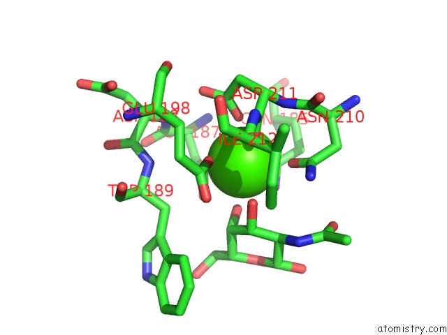
Mono view
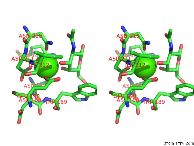
Stereo pair view
|
A full contact list of Calcium with other atoms in the Ca binding
site number 5 of Mannose-Binding Protein-A Mutant (Qpdwgh) Complexed with N- Acetyl-D-Galactosamine within 5.0Å range:
probe
|
atom
|
residue
|
distance (Å)
|
B
|
Occ
|
2:Ca2
b:35.7
occ:1.00
|
OE1
|
2:GLU198
|
2.2
|
40.8
|
1.0
|
OD1
|
2:ASP211
|
2.4
|
30.9
|
1.0
|
OE1
|
2:GLN185
|
2.4
|
44.3
|
1.0
|
OD1
|
2:ASP187
|
2.4
|
41.2
|
1.0
|
O
|
2:ASP211
|
2.5
|
33.9
|
1.0
|
OD1
|
2:ASN210
|
2.5
|
41.9
|
1.0
|
O3
|
2:NGA1
|
2.6
|
39.7
|
1.0
|
O4
|
2:NGA1
|
2.7
|
40.6
|
1.0
|
CD
|
2:GLU198
|
3.3
|
40.4
|
1.0
|
CD
|
2:GLN185
|
3.3
|
42.8
|
1.0
|
CG
|
2:ASP211
|
3.4
|
35.1
|
1.0
|
C4
|
2:NGA1
|
3.5
|
43.7
|
1.0
|
CG
|
2:ASP187
|
3.5
|
42.7
|
1.0
|
CG
|
2:ASN210
|
3.5
|
41.5
|
1.0
|
C3
|
2:NGA1
|
3.5
|
42.4
|
1.0
|
C
|
2:ASP211
|
3.5
|
36.1
|
1.0
|
OE2
|
2:GLU198
|
3.7
|
41.0
|
1.0
|
N
|
2:ASP211
|
3.7
|
36.1
|
1.0
|
NE2
|
2:GLN185
|
3.8
|
43.2
|
1.0
|
ND2
|
2:ASN210
|
3.9
|
40.5
|
1.0
|
OD2
|
2:ASP187
|
3.9
|
41.8
|
1.0
|
CA
|
2:ASP211
|
4.1
|
35.3
|
1.0
|
OD2
|
2:ASP211
|
4.1
|
32.9
|
1.0
|
C2
|
2:NGA1
|
4.2
|
43.4
|
1.0
|
CB
|
2:ASP211
|
4.3
|
34.7
|
1.0
|
N
|
2:ASP187
|
4.3
|
41.6
|
1.0
|
CG
|
2:GLN185
|
4.5
|
42.6
|
1.0
|
CB
|
2:TRP189
|
4.5
|
51.2
|
1.0
|
CG
|
2:GLU198
|
4.6
|
39.9
|
1.0
|
C
|
2:ASN210
|
4.6
|
38.5
|
1.0
|
N
|
2:ILE212
|
4.7
|
35.5
|
1.0
|
CB
|
2:GLU198
|
4.7
|
40.3
|
1.0
|
CB
|
2:ASP187
|
4.7
|
41.4
|
1.0
|
N
|
2:TRP189
|
4.7
|
46.6
|
1.0
|
N
|
2:ASP188
|
4.8
|
41.6
|
1.0
|
CB
|
2:ASN210
|
4.8
|
39.8
|
1.0
|
C5
|
2:NGA1
|
4.9
|
45.7
|
1.0
|
CA
|
2:ASP187
|
4.9
|
41.3
|
1.0
|
CA
|
2:ASN210
|
5.0
|
39.3
|
1.0
|
|
Calcium binding site 6 out
of 9 in 1bch
Go back to
Calcium Binding Sites List in 1bch
Calcium binding site 6 out
of 9 in the Mannose-Binding Protein-A Mutant (Qpdwgh) Complexed with N- Acetyl-D-Galactosamine
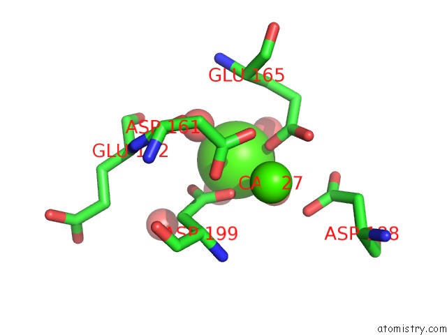
Mono view
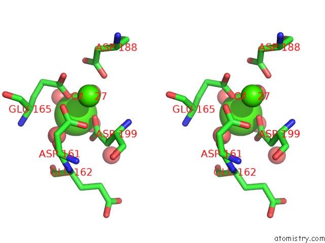
Stereo pair view
|
A full contact list of Calcium with other atoms in the Ca binding
site number 6 of Mannose-Binding Protein-A Mutant (Qpdwgh) Complexed with N- Acetyl-D-Galactosamine within 5.0Å range:
probe
|
atom
|
residue
|
distance (Å)
|
B
|
Occ
|
2:Ca3
b:48.5
occ:1.00
|
OE1
|
2:GLU165
|
2.3
|
37.9
|
1.0
|
O
|
2:HOH293
|
2.3
|
38.8
|
1.0
|
O
|
2:HOH312
|
2.4
|
45.4
|
1.0
|
OD1
|
2:ASP199
|
2.4
|
39.0
|
1.0
|
OD2
|
2:ASP199
|
2.5
|
39.6
|
1.0
|
O
|
2:HOH310
|
2.5
|
40.1
|
1.0
|
O
|
2:HOH311
|
2.6
|
41.4
|
1.0
|
CG
|
2:ASP199
|
2.8
|
37.9
|
1.0
|
CD
|
2:GLU165
|
3.4
|
34.7
|
1.0
|
CB
|
2:GLU165
|
3.9
|
39.9
|
1.0
|
CA
|
2:CA227
|
4.0
|
34.1
|
1.0
|
CG
|
2:GLU165
|
4.1
|
37.7
|
1.0
|
O
|
2:ASP161
|
4.2
|
38.3
|
1.0
|
CB
|
2:ASP199
|
4.3
|
37.6
|
1.0
|
OD1
|
2:ASP188
|
4.3
|
41.0
|
1.0
|
CA
|
2:GLU165
|
4.4
|
39.6
|
1.0
|
OE2
|
2:GLU165
|
4.4
|
30.7
|
1.0
|
OD2
|
2:ASP188
|
4.6
|
43.3
|
1.0
|
O
|
2:HOH271
|
4.7
|
50.8
|
1.0
|
OD2
|
2:ASP161
|
4.8
|
32.7
|
1.0
|
O
|
2:GLU162
|
4.8
|
43.8
|
1.0
|
CG
|
2:ASP188
|
4.9
|
43.4
|
1.0
|
N
|
2:GLU165
|
4.9
|
40.4
|
1.0
|
OD1
|
2:ASP161
|
5.0
|
32.5
|
1.0
|
|
Calcium binding site 7 out
of 9 in 1bch
Go back to
Calcium Binding Sites List in 1bch
Calcium binding site 7 out
of 9 in the Mannose-Binding Protein-A Mutant (Qpdwgh) Complexed with N- Acetyl-D-Galactosamine
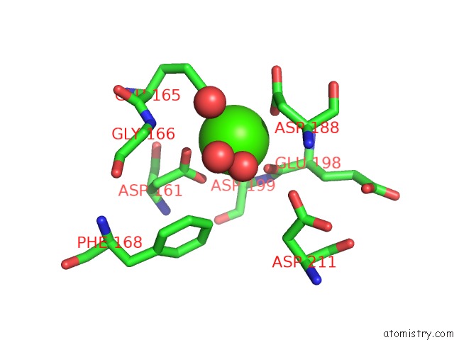
Mono view
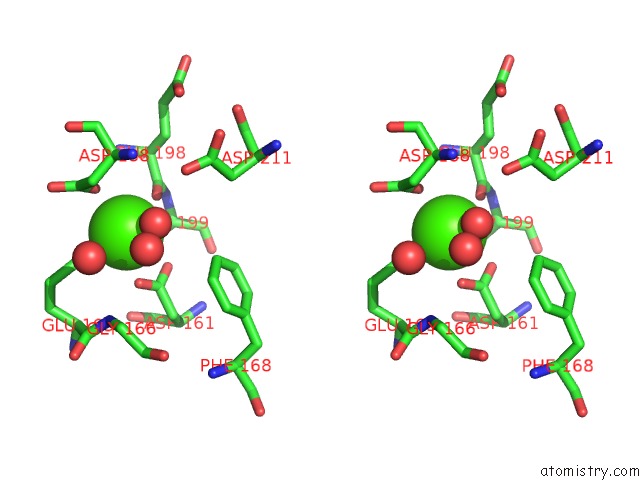
Stereo pair view
|
A full contact list of Calcium with other atoms in the Ca binding
site number 7 of Mannose-Binding Protein-A Mutant (Qpdwgh) Complexed with N- Acetyl-D-Galactosamine within 5.0Å range:
probe
|
atom
|
residue
|
distance (Å)
|
B
|
Occ
|
3:Ca227
b:27.9
occ:1.00
|
O
|
3:GLU198
|
2.3
|
28.0
|
1.0
|
O
|
3:HOH242
|
2.4
|
27.1
|
1.0
|
OD1
|
3:ASP199
|
2.4
|
23.9
|
1.0
|
OE1
|
3:GLU165
|
2.5
|
23.8
|
1.0
|
OD2
|
3:ASP161
|
2.5
|
33.1
|
1.0
|
OD1
|
3:ASP188
|
2.6
|
36.0
|
1.0
|
OE2
|
3:GLU165
|
2.7
|
25.5
|
1.0
|
OD1
|
3:ASP161
|
2.8
|
31.2
|
1.0
|
CD
|
3:GLU165
|
2.9
|
25.7
|
1.0
|
CG
|
3:ASP161
|
3.0
|
32.9
|
1.0
|
C
|
3:GLU198
|
3.2
|
27.4
|
1.0
|
CG
|
3:ASP188
|
3.3
|
36.0
|
1.0
|
CG
|
3:ASP199
|
3.5
|
25.3
|
1.0
|
CA
|
3:ASP199
|
3.7
|
27.7
|
1.0
|
CB
|
3:ASP188
|
3.7
|
36.6
|
1.0
|
N
|
3:ASP199
|
3.8
|
26.2
|
1.0
|
CB
|
3:ASP199
|
4.2
|
27.0
|
1.0
|
OD2
|
3:ASP188
|
4.2
|
34.6
|
1.0
|
N
|
3:GLU198
|
4.3
|
29.5
|
1.0
|
CA
|
3:GLU198
|
4.3
|
28.9
|
1.0
|
CA
|
3:ASP188
|
4.4
|
38.3
|
1.0
|
OD2
|
3:ASP199
|
4.4
|
24.6
|
1.0
|
CG
|
3:GLU165
|
4.4
|
28.4
|
1.0
|
CB
|
3:ASP161
|
4.5
|
32.2
|
1.0
|
N
|
3:GLY166
|
4.6
|
35.6
|
1.0
|
CZ
|
3:PHE168
|
4.7
|
36.1
|
1.0
|
O
|
3:HOH276
|
4.7
|
37.7
|
1.0
|
O
|
3:ASP161
|
4.8
|
33.5
|
1.0
|
O
|
3:HOH244
|
4.8
|
36.8
|
1.0
|
OD2
|
3:ASP211
|
4.8
|
32.3
|
1.0
|
CA
|
3:GLU165
|
4.9
|
32.2
|
1.0
|
C
|
3:ASP199
|
4.9
|
28.4
|
1.0
|
|
Calcium binding site 8 out
of 9 in 1bch
Go back to
Calcium Binding Sites List in 1bch
Calcium binding site 8 out
of 9 in the Mannose-Binding Protein-A Mutant (Qpdwgh) Complexed with N- Acetyl-D-Galactosamine
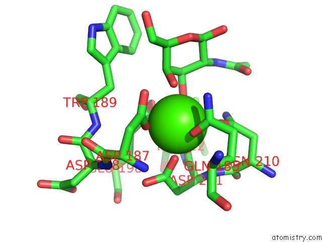
Mono view
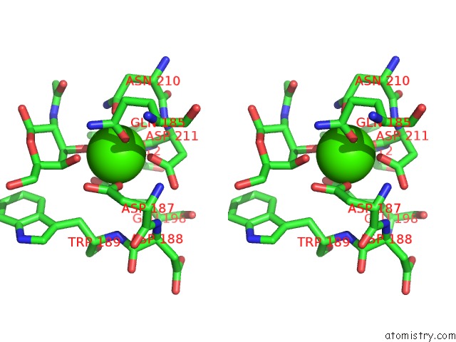
Stereo pair view
|
A full contact list of Calcium with other atoms in the Ca binding
site number 8 of Mannose-Binding Protein-A Mutant (Qpdwgh) Complexed with N- Acetyl-D-Galactosamine within 5.0Å range:
probe
|
atom
|
residue
|
distance (Å)
|
B
|
Occ
|
3:Ca2
b:35.1
occ:1.00
|
OD1
|
3:ASP211
|
2.1
|
29.9
|
1.0
|
OE1
|
3:GLN185
|
2.4
|
43.7
|
1.0
|
OE1
|
3:GLU198
|
2.4
|
30.8
|
1.0
|
O3
|
3:NGA1
|
2.5
|
43.2
|
1.0
|
OD1
|
3:ASP187
|
2.5
|
38.5
|
1.0
|
O
|
3:ASP211
|
2.5
|
28.7
|
1.0
|
OD1
|
3:ASN210
|
2.6
|
31.7
|
1.0
|
O4
|
3:NGA1
|
2.6
|
44.2
|
1.0
|
CD
|
3:GLN185
|
3.2
|
43.7
|
1.0
|
CG
|
3:ASP211
|
3.3
|
32.4
|
1.0
|
C4
|
3:NGA1
|
3.4
|
44.6
|
1.0
|
CD
|
3:GLU198
|
3.4
|
29.8
|
1.0
|
C3
|
3:NGA1
|
3.4
|
45.7
|
1.0
|
CG
|
3:ASP187
|
3.5
|
40.7
|
1.0
|
CG
|
3:ASN210
|
3.5
|
33.1
|
1.0
|
C
|
3:ASP211
|
3.6
|
28.6
|
1.0
|
NE2
|
3:GLN185
|
3.6
|
43.1
|
1.0
|
OE2
|
3:GLU198
|
3.8
|
30.0
|
1.0
|
N
|
3:ASP211
|
3.8
|
30.9
|
1.0
|
ND2
|
3:ASN210
|
3.8
|
28.9
|
1.0
|
OD2
|
3:ASP187
|
3.9
|
39.9
|
1.0
|
OD2
|
3:ASP211
|
4.0
|
32.3
|
1.0
|
CA
|
3:ASP211
|
4.1
|
29.7
|
1.0
|
C2
|
3:NGA1
|
4.2
|
47.1
|
1.0
|
CB
|
3:ASP211
|
4.3
|
30.8
|
1.0
|
N
|
3:ASP187
|
4.3
|
40.6
|
1.0
|
CG
|
3:GLN185
|
4.5
|
43.4
|
1.0
|
CB
|
3:TRP189
|
4.7
|
39.1
|
1.0
|
CG
|
3:GLU198
|
4.7
|
29.2
|
1.0
|
C
|
3:ASN210
|
4.7
|
32.8
|
1.0
|
CB
|
3:ASP187
|
4.7
|
40.7
|
1.0
|
N
|
3:ILE212
|
4.8
|
27.4
|
1.0
|
C5
|
3:NGA1
|
4.8
|
47.2
|
1.0
|
CB
|
3:GLU198
|
4.8
|
27.6
|
1.0
|
CB
|
3:ASN210
|
4.8
|
31.9
|
1.0
|
N
|
3:TRP189
|
4.9
|
37.6
|
1.0
|
N
|
3:ASP188
|
4.9
|
38.9
|
1.0
|
CA
|
3:ASP187
|
5.0
|
41.4
|
1.0
|
|
Calcium binding site 9 out
of 9 in 1bch
Go back to
Calcium Binding Sites List in 1bch
Calcium binding site 9 out
of 9 in the Mannose-Binding Protein-A Mutant (Qpdwgh) Complexed with N- Acetyl-D-Galactosamine
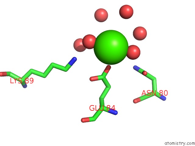
Mono view
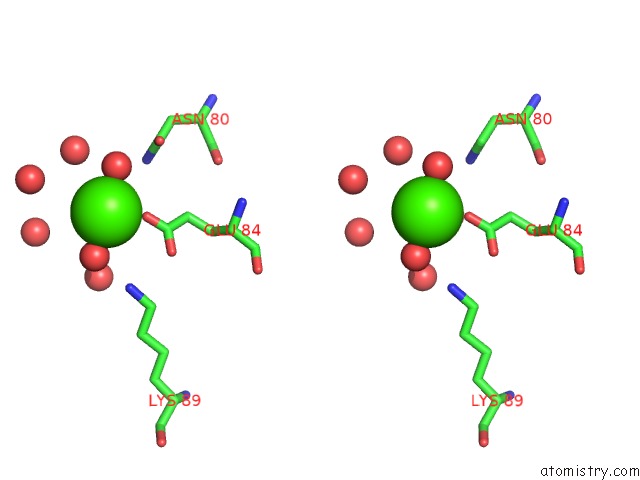
Stereo pair view
|
A full contact list of Calcium with other atoms in the Ca binding
site number 9 of Mannose-Binding Protein-A Mutant (Qpdwgh) Complexed with N- Acetyl-D-Galactosamine within 5.0Å range:
probe
|
atom
|
residue
|
distance (Å)
|
B
|
Occ
|
3:Ca3
b:27.8
occ:1.00
|
O
|
1:HOH239
|
2.3
|
31.6
|
1.0
|
O
|
3:HOH228
|
2.3
|
24.0
|
1.0
|
OE2
|
3:GLU84
|
2.4
|
25.2
|
1.0
|
O
|
3:HOH249
|
2.4
|
26.9
|
1.0
|
CD
|
3:GLU84
|
3.5
|
23.6
|
1.0
|
OE1
|
3:GLU84
|
4.0
|
28.0
|
1.0
|
NZ
|
1:LYS89
|
4.2
|
26.9
|
1.0
|
O
|
3:HOH236
|
4.4
|
32.9
|
1.0
|
O
|
3:HOH290
|
4.6
|
46.5
|
1.0
|
ND2
|
3:ASN80
|
4.6
|
27.8
|
1.0
|
OD1
|
3:ASN80
|
4.6
|
25.6
|
1.0
|
O
|
1:HOH271
|
4.7
|
34.9
|
1.0
|
O
|
3:HOH248
|
4.7
|
38.2
|
1.0
|
CG
|
3:GLU84
|
4.8
|
21.3
|
1.0
|
O
|
3:HOH296
|
4.9
|
54.5
|
1.0
|
|
Reference:
A.R.Kolatkar,
A.K.Leung,
R.Isecke,
R.Brossmer,
K.Drickamer,
W.I.Weis.
Mechanism of N-Acetylgalactosamine Binding to A C-Type Animal Lectin Carbohydrate-Recognition Domain. J.Biol.Chem. V. 273 19502 1998.
ISSN: ISSN 0021-9258
PubMed: 9677372
DOI: 10.1074/JBC.273.31.19502
Page generated: Thu Jul 11 06:20:26 2024
|