Calcium »
PDB 1b85-1bjj »
1be6 »
Calcium in PDB 1be6: Trans-Cinnamoyl-Subtilisin in Anhydrous Acetonitrile
Enzymatic activity of Trans-Cinnamoyl-Subtilisin in Anhydrous Acetonitrile
All present enzymatic activity of Trans-Cinnamoyl-Subtilisin in Anhydrous Acetonitrile:
3.4.21.62;
3.4.21.62;
Protein crystallography data
The structure of Trans-Cinnamoyl-Subtilisin in Anhydrous Acetonitrile, PDB code: 1be6
was solved by
J.L.Schmitke,
L.J.Stern,
A.M.Klibanov,
with X-Ray Crystallography technique. A brief refinement statistics is given in the table below:
| Resolution Low / High (Å) | 8.00 / 2.15 |
| Space group | P 21 21 21 |
| Cell size a, b, c (Å), α, β, γ (°) | 76.409, 55.261, 52.824, 90.00, 90.00, 90.00 |
| R / Rfree (%) | 19.7 / 23.4 |
Calcium Binding Sites:
The binding sites of Calcium atom in the Trans-Cinnamoyl-Subtilisin in Anhydrous Acetonitrile
(pdb code 1be6). This binding sites where shown within
5.0 Angstroms radius around Calcium atom.
In total only one binding site of Calcium was determined in the Trans-Cinnamoyl-Subtilisin in Anhydrous Acetonitrile, PDB code: 1be6:
In total only one binding site of Calcium was determined in the Trans-Cinnamoyl-Subtilisin in Anhydrous Acetonitrile, PDB code: 1be6:
Calcium binding site 1 out of 1 in 1be6
Go back to
Calcium binding site 1 out
of 1 in the Trans-Cinnamoyl-Subtilisin in Anhydrous Acetonitrile
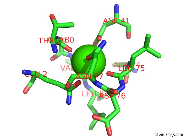
Mono view
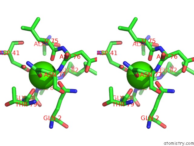
Stereo pair view
Mono view
Stereo pair view
A full contact list of Calcium with other atoms in the Ca binding
site number 1 of Trans-Cinnamoyl-Subtilisin in Anhydrous Acetonitrile within 5.0Å range:
|
Reference:
J.L.Schmitke,
L.J.Stern,
A.M.Klibanov.
Comparison of X-Ray Crystal Structures of An Acyl-Enzyme Intermediate of Subtilisin Carlsberg Formed in Anhydrous Acetonitrile and in Water. Proc.Natl.Acad.Sci.Usa V. 95 12918 1998.
ISSN: ISSN 0027-8424
PubMed: 9789015
DOI: 10.1073/PNAS.95.22.12918
Page generated: Mon Jul 7 13:41:25 2025
ISSN: ISSN 0027-8424
PubMed: 9789015
DOI: 10.1073/PNAS.95.22.12918
Last articles
Cl in 5JNYCl in 5JMS
Cl in 5JMG
Cl in 5JML
Cl in 5JKL
Cl in 5JMQ
Cl in 5JLC
Cl in 5JLJ
Cl in 5JKE
Cl in 5JKK