Calcium »
PDB 1b85-1bjj »
1bf2 »
Calcium in PDB 1bf2: Structure of Pseudomonas Isoamylase
Enzymatic activity of Structure of Pseudomonas Isoamylase
All present enzymatic activity of Structure of Pseudomonas Isoamylase:
3.2.1.68;
3.2.1.68;
Protein crystallography data
The structure of Structure of Pseudomonas Isoamylase, PDB code: 1bf2
was solved by
Y.Katsuya,
Y.Mezaki,
M.Kubota,
Y.Matsuura,
with X-Ray Crystallography technique. A brief refinement statistics is given in the table below:
| Resolution Low / High (Å) | 10.00 / 2.00 |
| Space group | P 21 21 21 |
| Cell size a, b, c (Å), α, β, γ (°) | 138.900, 152.400, 53.500, 90.00, 90.00, 90.00 |
| R / Rfree (%) | 16.1 / 21.4 |
Calcium Binding Sites:
The binding sites of Calcium atom in the Structure of Pseudomonas Isoamylase
(pdb code 1bf2). This binding sites where shown within
5.0 Angstroms radius around Calcium atom.
In total only one binding site of Calcium was determined in the Structure of Pseudomonas Isoamylase, PDB code: 1bf2:
In total only one binding site of Calcium was determined in the Structure of Pseudomonas Isoamylase, PDB code: 1bf2:
Calcium binding site 1 out of 1 in 1bf2
Go back to
Calcium binding site 1 out
of 1 in the Structure of Pseudomonas Isoamylase
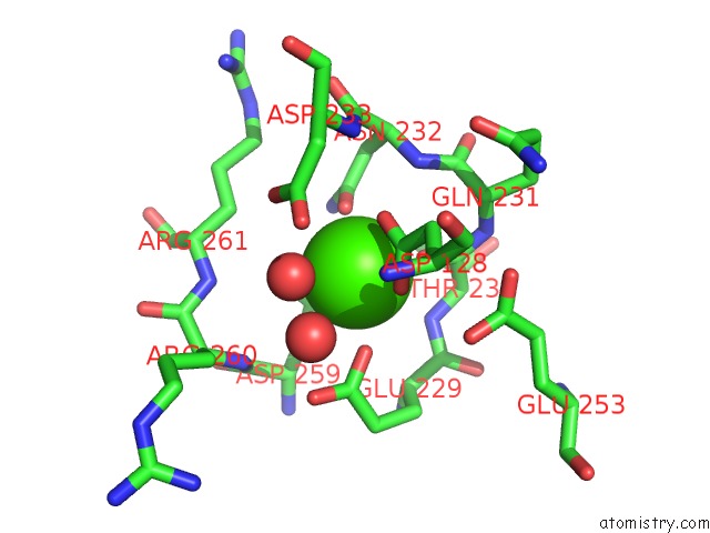
Mono view
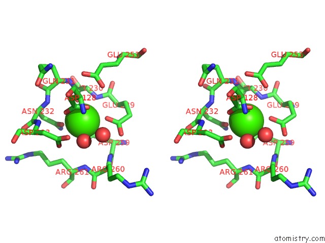
Stereo pair view
Mono view
Stereo pair view
A full contact list of Calcium with other atoms in the Ca binding
site number 1 of Structure of Pseudomonas Isoamylase within 5.0Å range:
|
Reference:
Y.Katsuya,
Y.Mezaki,
M.Kubota,
Y.Matsuura.
Three-Dimensional Structure of Pseudomonas Isoamylase at 2.2 A Resolution. J.Mol.Biol. V. 281 885 1998.
ISSN: ISSN 0022-2836
PubMed: 9719642
DOI: 10.1006/JMBI.1998.1992
Page generated: Mon Jul 7 13:41:47 2025
ISSN: ISSN 0022-2836
PubMed: 9719642
DOI: 10.1006/JMBI.1998.1992
Last articles
Cl in 5I9XCl in 5I7O
Cl in 5I8P
Cl in 5I9L
Cl in 5I8A
Cl in 5I7H
Cl in 5I5X
Cl in 5I6X
Cl in 5I5Y
Cl in 5I6U