Calcium »
PDB 1b85-1bjj »
1bj3 »
Calcium in PDB 1bj3: Crystal Structure of Coagulation Factor IX-Binding Protein (IX-Bp) From Venom of Habu Snake with A Heterodimer of C-Type Lectin Domains
Protein crystallography data
The structure of Crystal Structure of Coagulation Factor IX-Binding Protein (IX-Bp) From Venom of Habu Snake with A Heterodimer of C-Type Lectin Domains, PDB code: 1bj3
was solved by
H.Mizuno,
Z.Fujimoto,
M.Koizumi,
H.Kano,
H.Atoda,
T.Morita,
with X-Ray Crystallography technique. A brief refinement statistics is given in the table below:
| Resolution Low / High (Å) | 6.00 / 2.60 |
| Space group | P 1 21 1 |
| Cell size a, b, c (Å), α, β, γ (°) | 58.569, 56.794, 39.616, 90.00, 95.80, 90.00 |
| R / Rfree (%) | 18.2 / 27.1 |
Calcium Binding Sites:
The binding sites of Calcium atom in the Crystal Structure of Coagulation Factor IX-Binding Protein (IX-Bp) From Venom of Habu Snake with A Heterodimer of C-Type Lectin Domains
(pdb code 1bj3). This binding sites where shown within
5.0 Angstroms radius around Calcium atom.
In total 2 binding sites of Calcium where determined in the Crystal Structure of Coagulation Factor IX-Binding Protein (IX-Bp) From Venom of Habu Snake with A Heterodimer of C-Type Lectin Domains, PDB code: 1bj3:
Jump to Calcium binding site number: 1; 2;
In total 2 binding sites of Calcium where determined in the Crystal Structure of Coagulation Factor IX-Binding Protein (IX-Bp) From Venom of Habu Snake with A Heterodimer of C-Type Lectin Domains, PDB code: 1bj3:
Jump to Calcium binding site number: 1; 2;
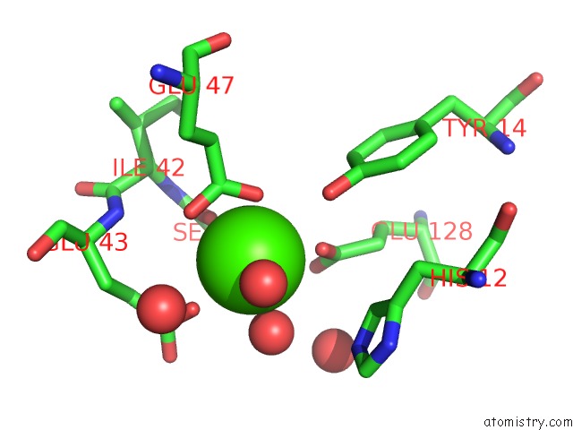
Calcium binding site 1 out of 2 in 1bj3
Go back to
Calcium binding site 1 out
of 2 in the Crystal Structure of Coagulation Factor IX-Binding Protein (IX-Bp) From Venom of Habu Snake with A Heterodimer of C-Type Lectin Domains
Mono view
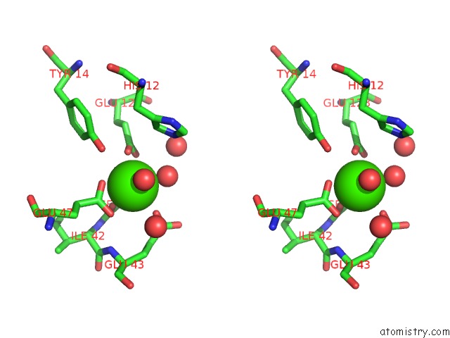
Stereo pair view
Mono view
Stereo pair view
A full contact list of Calcium with other atoms in the Ca binding
site number 1 of Crystal Structure of Coagulation Factor IX-Binding Protein (IX-Bp) From Venom of Habu Snake with A Heterodimer of C-Type Lectin Domains within 5.0Å range:
|
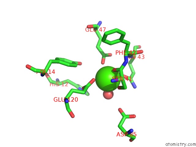
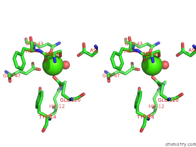
Calcium binding site 2 out of 2 in 1bj3
Go back to
Calcium binding site 2 out
of 2 in the Crystal Structure of Coagulation Factor IX-Binding Protein (IX-Bp) From Venom of Habu Snake with A Heterodimer of C-Type Lectin Domains
Mono view
Stereo pair view
Mono view
Stereo pair view
A full contact list of Calcium with other atoms in the Ca binding
site number 2 of Crystal Structure of Coagulation Factor IX-Binding Protein (IX-Bp) From Venom of Habu Snake with A Heterodimer of C-Type Lectin Domains within 5.0Å range:
|
Reference:
H.Mizuno,
Z.Fujimoto,
M.Koizumi,
H.Kano,
H.Atoda,
T.Morita.
Crystal Structure of Coagulation Factor IX-Binding Protein From Habu Snake Venom at 2.6 A: Implication of Central Loop Swapping Based on Deletion in the Linker Region. J.Mol.Biol. V. 289 103 1999.
ISSN: ISSN 0022-2836
PubMed: 10339409
DOI: 10.1006/JMBI.1999.2756
Page generated: Thu Jul 11 06:26:21 2024
ISSN: ISSN 0022-2836
PubMed: 10339409
DOI: 10.1006/JMBI.1999.2756
Last articles
Zn in 9J0NZn in 9J0O
Zn in 9J0P
Zn in 9FJX
Zn in 9EKB
Zn in 9C0F
Zn in 9CAH
Zn in 9CH0
Zn in 9CH3
Zn in 9CH1