Calcium »
PDB 1bjq-1byh »
1bsw »
Calcium in PDB 1bsw: Acutolysin A From Snake Venom of Agkistrodon Acutus at pH 7.5
Protein crystallography data
The structure of Acutolysin A From Snake Venom of Agkistrodon Acutus at pH 7.5, PDB code: 1bsw
was solved by
W.Gong,
X.Zhu,
S.Liu,
M.Teng,
L.Niu,
with X-Ray Crystallography technique. A brief refinement statistics is given in the table below:
| Resolution Low / High (Å) | 10.00 / 1.95 |
| Space group | P 43 21 2 |
| Cell size a, b, c (Å), α, β, γ (°) | 63.170, 63.170, 95.350, 90.00, 90.00, 90.00 |
| R / Rfree (%) | 17.1 / n/a |
Other elements in 1bsw:
The structure of Acutolysin A From Snake Venom of Agkistrodon Acutus at pH 7.5 also contains other interesting chemical elements:
| Zinc | (Zn) | 1 atom |
Calcium Binding Sites:
The binding sites of Calcium atom in the Acutolysin A From Snake Venom of Agkistrodon Acutus at pH 7.5
(pdb code 1bsw). This binding sites where shown within
5.0 Angstroms radius around Calcium atom.
In total only one binding site of Calcium was determined in the Acutolysin A From Snake Venom of Agkistrodon Acutus at pH 7.5, PDB code: 1bsw:
In total only one binding site of Calcium was determined in the Acutolysin A From Snake Venom of Agkistrodon Acutus at pH 7.5, PDB code: 1bsw:
Calcium binding site 1 out of 1 in 1bsw
Go back to
Calcium binding site 1 out
of 1 in the Acutolysin A From Snake Venom of Agkistrodon Acutus at pH 7.5
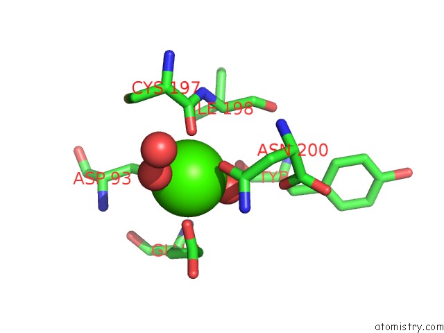
Mono view
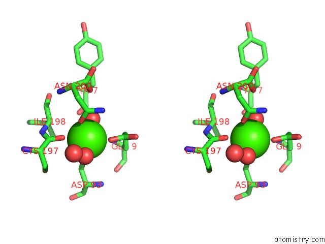
Stereo pair view
Mono view
Stereo pair view
A full contact list of Calcium with other atoms in the Ca binding
site number 1 of Acutolysin A From Snake Venom of Agkistrodon Acutus at pH 7.5 within 5.0Å range:
|
Reference:
W.Gong,
X.Zhu,
S.Liu,
M.Teng,
L.Niu.
Crystal Structures of Acutolysin A, A Three-Disulfide Hemorrhagic Zinc Metalloproteinase From the Snake Venom of Agkistrodon Acutus. J.Mol.Biol. V. 283 657 1998.
ISSN: ISSN 0022-2836
PubMed: 9784374
DOI: 10.1006/JMBI.1998.2110
Page generated: Mon Jul 7 13:48:13 2025
ISSN: ISSN 0022-2836
PubMed: 9784374
DOI: 10.1006/JMBI.1998.2110
Last articles
Ca in 7NM3Ca in 7NLM
Ca in 7NM1
Ca in 7NL7
Ca in 7NL6
Ca in 7NLK
Ca in 7NLA
Ca in 7NK5
Ca in 7NK3
Ca in 7NJ8