Calcium »
PDB 1byn-1c5v »
1bzx »
Calcium in PDB 1bzx: The Crystal Structure of Anionic Salmon Trypsin in Complex with Bovine Pancreatic Trypsin Inhibitor
Enzymatic activity of The Crystal Structure of Anionic Salmon Trypsin in Complex with Bovine Pancreatic Trypsin Inhibitor
All present enzymatic activity of The Crystal Structure of Anionic Salmon Trypsin in Complex with Bovine Pancreatic Trypsin Inhibitor:
3.4.21.4;
3.4.21.4;
Protein crystallography data
The structure of The Crystal Structure of Anionic Salmon Trypsin in Complex with Bovine Pancreatic Trypsin Inhibitor, PDB code: 1bzx
was solved by
R.Helland,
I.Leiros,
G.I.Berglund,
N.P.Willassen,
A.O.Smalas,
with X-Ray Crystallography technique. A brief refinement statistics is given in the table below:
| Resolution Low / High (Å) | 8.00 / 2.10 |
| Space group | P 61 2 2 |
| Cell size a, b, c (Å), α, β, γ (°) | 84.120, 84.120, 222.150, 90.00, 90.00, 120.00 |
| R / Rfree (%) | 20.6 / 23.8 |
Calcium Binding Sites:
The binding sites of Calcium atom in the The Crystal Structure of Anionic Salmon Trypsin in Complex with Bovine Pancreatic Trypsin Inhibitor
(pdb code 1bzx). This binding sites where shown within
5.0 Angstroms radius around Calcium atom.
In total only one binding site of Calcium was determined in the The Crystal Structure of Anionic Salmon Trypsin in Complex with Bovine Pancreatic Trypsin Inhibitor, PDB code: 1bzx:
In total only one binding site of Calcium was determined in the The Crystal Structure of Anionic Salmon Trypsin in Complex with Bovine Pancreatic Trypsin Inhibitor, PDB code: 1bzx:
Calcium binding site 1 out of 1 in 1bzx
Go back to
Calcium binding site 1 out
of 1 in the The Crystal Structure of Anionic Salmon Trypsin in Complex with Bovine Pancreatic Trypsin Inhibitor
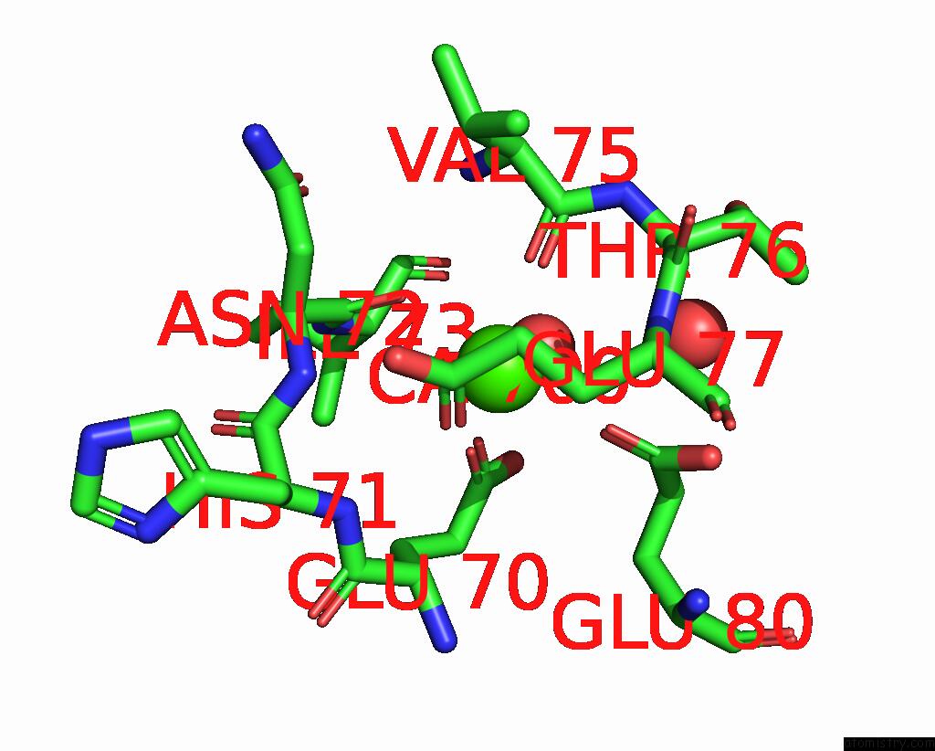
Mono view
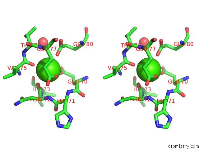
Stereo pair view
Mono view
Stereo pair view
A full contact list of Calcium with other atoms in the Ca binding
site number 1 of The Crystal Structure of Anionic Salmon Trypsin in Complex with Bovine Pancreatic Trypsin Inhibitor within 5.0Å range:
|
Reference:
R.Helland,
I.Leiros,
G.I.Berglund,
N.P.Willassen,
A.O.Smalas.
The Crystal Structure of Anionic Salmon Trypsin in Complex with Bovine Pancreatic Trypsin Inhibitor. Eur.J.Biochem. V. 256 317 1998.
ISSN: ISSN 0014-2956
PubMed: 9760170
DOI: 10.1046/J.1432-1327.1998.2560317.X
Page generated: Thu Jul 11 06:46:49 2024
ISSN: ISSN 0014-2956
PubMed: 9760170
DOI: 10.1046/J.1432-1327.1998.2560317.X
Last articles
Zn in 9MJ5Zn in 9HNW
Zn in 9G0L
Zn in 9FNE
Zn in 9DZN
Zn in 9E0I
Zn in 9D32
Zn in 9DAK
Zn in 8ZXC
Zn in 8ZUF