Calcium »
PDB 1c74-1cjy »
1cdm »
Calcium in PDB 1cdm: Modulation of Calmodulin Plasticity in Molecular Recognition on the Basis of X-Ray Structures
Protein crystallography data
The structure of Modulation of Calmodulin Plasticity in Molecular Recognition on the Basis of X-Ray Structures, PDB code: 1cdm
was solved by
W.E.Meador,
F.A.Quiocho,
with X-Ray Crystallography technique. A brief refinement statistics is given in the table below:
| Resolution Low / High (Å) | 10.00 / 2.00 |
| Space group | C 2 2 21 |
| Cell size a, b, c (Å), α, β, γ (°) | 39.000, 75.200, 120.150, 90.00, 90.00, 90.00 |
| R / Rfree (%) | 20.8 / n/a |
Calcium Binding Sites:
The binding sites of Calcium atom in the Modulation of Calmodulin Plasticity in Molecular Recognition on the Basis of X-Ray Structures
(pdb code 1cdm). This binding sites where shown within
5.0 Angstroms radius around Calcium atom.
In total 4 binding sites of Calcium where determined in the Modulation of Calmodulin Plasticity in Molecular Recognition on the Basis of X-Ray Structures, PDB code: 1cdm:
Jump to Calcium binding site number: 1; 2; 3; 4;
In total 4 binding sites of Calcium where determined in the Modulation of Calmodulin Plasticity in Molecular Recognition on the Basis of X-Ray Structures, PDB code: 1cdm:
Jump to Calcium binding site number: 1; 2; 3; 4;
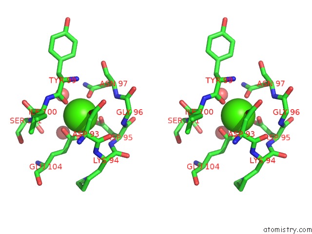
Calcium binding site 1 out of 4 in 1cdm
Go back to
Calcium binding site 1 out
of 4 in the Modulation of Calmodulin Plasticity in Molecular Recognition on the Basis of X-Ray Structures
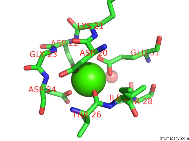
Mono view
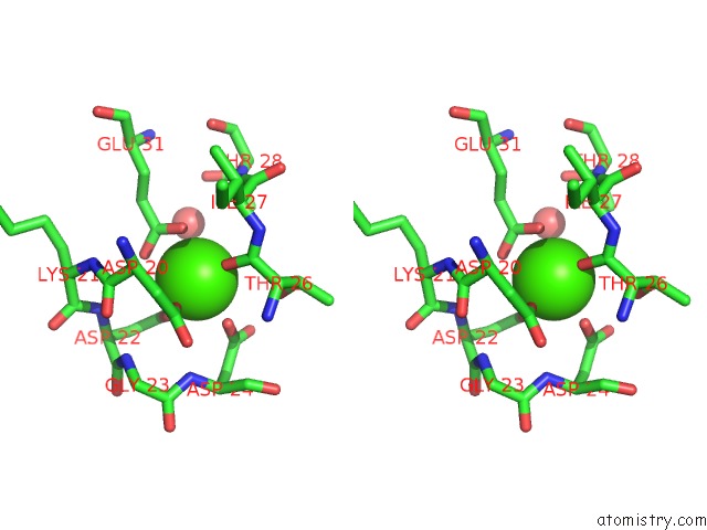
Stereo pair view
Mono view
Stereo pair view
A full contact list of Calcium with other atoms in the Ca binding
site number 1 of Modulation of Calmodulin Plasticity in Molecular Recognition on the Basis of X-Ray Structures within 5.0Å range:
|
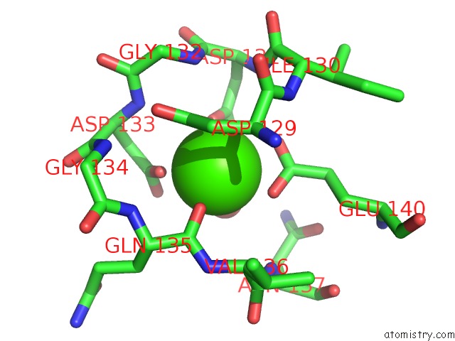
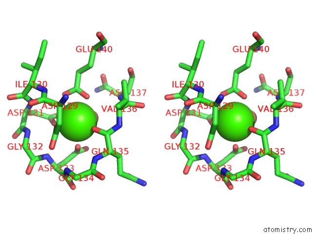
Calcium binding site 2 out of 4 in 1cdm
Go back to
Calcium binding site 2 out
of 4 in the Modulation of Calmodulin Plasticity in Molecular Recognition on the Basis of X-Ray Structures
Mono view
Stereo pair view
Mono view
Stereo pair view
A full contact list of Calcium with other atoms in the Ca binding
site number 2 of Modulation of Calmodulin Plasticity in Molecular Recognition on the Basis of X-Ray Structures within 5.0Å range:
|
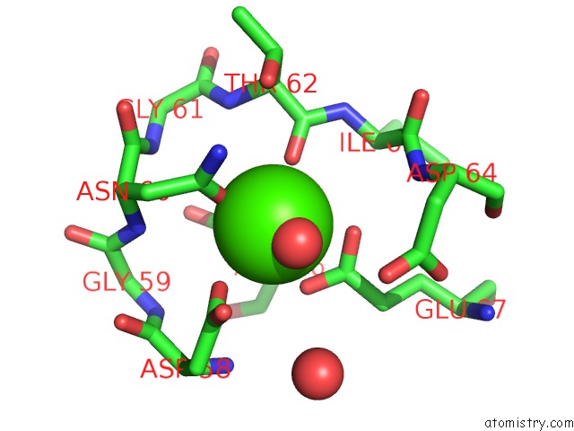
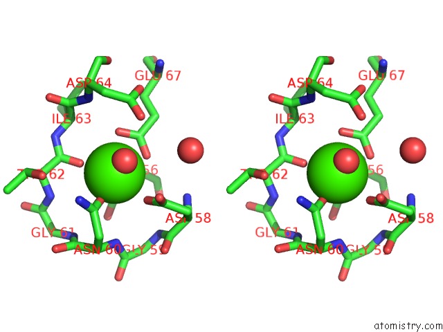
Calcium binding site 3 out of 4 in 1cdm
Go back to
Calcium binding site 3 out
of 4 in the Modulation of Calmodulin Plasticity in Molecular Recognition on the Basis of X-Ray Structures

Mono view
Stereo pair view
Mono view
Stereo pair view
A full contact list of Calcium with other atoms in the Ca binding
site number 3 of Modulation of Calmodulin Plasticity in Molecular Recognition on the Basis of X-Ray Structures within 5.0Å range:
|
Calcium binding site 4 out of 4 in 1cdm
Go back to
Calcium binding site 4 out
of 4 in the Modulation of Calmodulin Plasticity in Molecular Recognition on the Basis of X-Ray Structures
Mono view
Stereo pair view
Mono view
Stereo pair view
A full contact list of Calcium with other atoms in the Ca binding
site number 4 of Modulation of Calmodulin Plasticity in Molecular Recognition on the Basis of X-Ray Structures within 5.0Å range:
|
Reference:
W.E.Meador,
A.R.Means,
F.A.Quiocho.
Modulation of Calmodulin Plasticity in Molecular Recognition on the Basis of X-Ray Structures. Science V. 262 1718 1993.
ISSN: ISSN 0036-8075
PubMed: 8259515
Page generated: Mon Jul 7 13:57:13 2025
ISSN: ISSN 0036-8075
PubMed: 8259515
Last articles
Cl in 5RAQCl in 5RAP
Cl in 5RAO
Cl in 5RAN
Cl in 5RAJ
Cl in 5RAM
Cl in 5RAL
Cl in 5RAK
Cl in 5RAF
Cl in 5RAG