Calcium »
PDB 1c74-1cjy »
1cgu »
Calcium in PDB 1cgu: Catalytic Center of Cyclodextrin Glycosyltransferase Derived From X-Ray Structure Analysis Combined with Site- Directed Mutagenesis
Enzymatic activity of Catalytic Center of Cyclodextrin Glycosyltransferase Derived From X-Ray Structure Analysis Combined with Site- Directed Mutagenesis
All present enzymatic activity of Catalytic Center of Cyclodextrin Glycosyltransferase Derived From X-Ray Structure Analysis Combined with Site- Directed Mutagenesis:
2.4.1.19;
2.4.1.19;
Protein crystallography data
The structure of Catalytic Center of Cyclodextrin Glycosyltransferase Derived From X-Ray Structure Analysis Combined with Site- Directed Mutagenesis, PDB code: 1cgu
was solved by
C.Klein,
J.Hollender,
H.Bender,
G.E.Schulz,
with X-Ray Crystallography technique. A brief refinement statistics is given in the table below:
| Resolution Low / High (Å) | 10.00 / 2.50 |
| Space group | P 21 21 21 |
| Cell size a, b, c (Å), α, β, γ (°) | 94.100, 105.000, 113.900, 90.00, 90.00, 90.00 |
| R / Rfree (%) | 18.7 / n/a |
Calcium Binding Sites:
The binding sites of Calcium atom in the Catalytic Center of Cyclodextrin Glycosyltransferase Derived From X-Ray Structure Analysis Combined with Site- Directed Mutagenesis
(pdb code 1cgu). This binding sites where shown within
5.0 Angstroms radius around Calcium atom.
In total 2 binding sites of Calcium where determined in the Catalytic Center of Cyclodextrin Glycosyltransferase Derived From X-Ray Structure Analysis Combined with Site- Directed Mutagenesis, PDB code: 1cgu:
Jump to Calcium binding site number: 1; 2;
In total 2 binding sites of Calcium where determined in the Catalytic Center of Cyclodextrin Glycosyltransferase Derived From X-Ray Structure Analysis Combined with Site- Directed Mutagenesis, PDB code: 1cgu:
Jump to Calcium binding site number: 1; 2;
Calcium binding site 1 out of 2 in 1cgu
Go back to
Calcium binding site 1 out
of 2 in the Catalytic Center of Cyclodextrin Glycosyltransferase Derived From X-Ray Structure Analysis Combined with Site- Directed Mutagenesis

Mono view
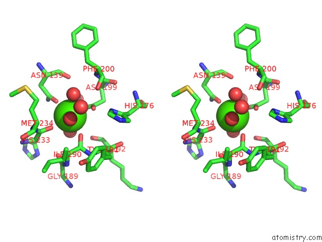
Stereo pair view
Mono view
Stereo pair view
A full contact list of Calcium with other atoms in the Ca binding
site number 1 of Catalytic Center of Cyclodextrin Glycosyltransferase Derived From X-Ray Structure Analysis Combined with Site- Directed Mutagenesis within 5.0Å range:
|
Calcium binding site 2 out of 2 in 1cgu
Go back to
Calcium binding site 2 out
of 2 in the Catalytic Center of Cyclodextrin Glycosyltransferase Derived From X-Ray Structure Analysis Combined with Site- Directed Mutagenesis

Mono view

Stereo pair view
Mono view
Stereo pair view
A full contact list of Calcium with other atoms in the Ca binding
site number 2 of Catalytic Center of Cyclodextrin Glycosyltransferase Derived From X-Ray Structure Analysis Combined with Site- Directed Mutagenesis within 5.0Å range:
|
Reference:
C.Klein,
J.Hollender,
H.Bender,
G.E.Schulz.
Catalytic Center of Cyclodextrin Glycosyltransferase Derived From X-Ray Structure Analysis Combined with Site-Directed Mutagenesis. Biochemistry V. 31 8740 1992.
ISSN: ISSN 0006-2960
PubMed: 1390660
DOI: 10.1021/BI00152A009
Page generated: Thu Jul 11 06:58:26 2024
ISSN: ISSN 0006-2960
PubMed: 1390660
DOI: 10.1021/BI00152A009
Last articles
Zn in 9MJ5Zn in 9HNW
Zn in 9G0L
Zn in 9FNE
Zn in 9DZN
Zn in 9E0I
Zn in 9D32
Zn in 9DAK
Zn in 8ZXC
Zn in 8ZUF