Calcium »
PDB 1ck6-1cxi »
1ck7 »
Calcium in PDB 1ck7: Gelatinase A (Full-Length)
Enzymatic activity of Gelatinase A (Full-Length)
All present enzymatic activity of Gelatinase A (Full-Length):
3.4.24.24;
3.4.24.24;
Protein crystallography data
The structure of Gelatinase A (Full-Length), PDB code: 1ck7
was solved by
E.Morgunova,
A.Tuuttila,
U.Bergmann,
M.Isupov,
Y.Lindqvist,
G.Schneider,
K.Tryggvason,
with X-Ray Crystallography technique. A brief refinement statistics is given in the table below:
| Resolution Low / High (Å) | 34.00 / 2.80 |
| Space group | I 41 2 2 |
| Cell size a, b, c (Å), α, β, γ (°) | 121.320, 121.320, 345.110, 90.00, 90.00, 90.00 |
| R / Rfree (%) | 28.6 / 32.7 |
Other elements in 1ck7:
The structure of Gelatinase A (Full-Length) also contains other interesting chemical elements:
| Zinc | (Zn) | 2 atoms |
| Chlorine | (Cl) | 1 atom |
| Sodium | (Na) | 1 atom |
Calcium Binding Sites:
The binding sites of Calcium atom in the Gelatinase A (Full-Length)
(pdb code 1ck7). This binding sites where shown within
5.0 Angstroms radius around Calcium atom.
In total 3 binding sites of Calcium where determined in the Gelatinase A (Full-Length), PDB code: 1ck7:
Jump to Calcium binding site number: 1; 2; 3;
In total 3 binding sites of Calcium where determined in the Gelatinase A (Full-Length), PDB code: 1ck7:
Jump to Calcium binding site number: 1; 2; 3;
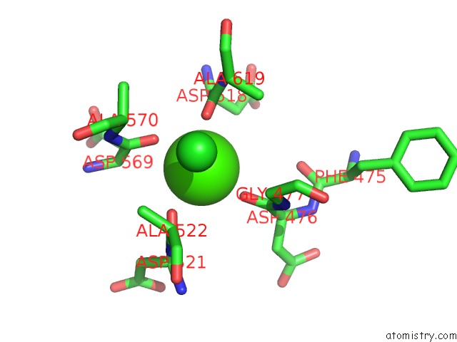
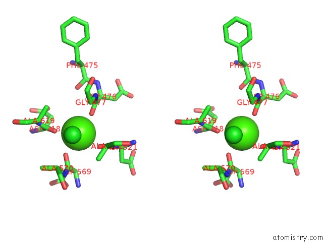
Calcium binding site 1 out of 3 in 1ck7
Go back to
Calcium binding site 1 out
of 3 in the Gelatinase A (Full-Length)
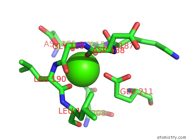
Mono view
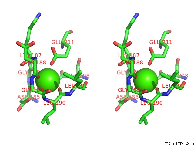
Stereo pair view
Mono view
Stereo pair view
A full contact list of Calcium with other atoms in the Ca binding
site number 1 of Gelatinase A (Full-Length) within 5.0Å range:
|
Calcium binding site 2 out of 3 in 1ck7
Go back to
Calcium binding site 2 out
of 3 in the Gelatinase A (Full-Length)
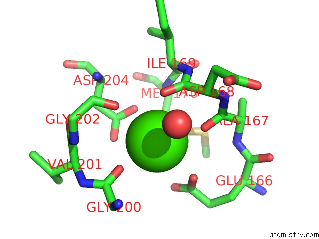
Mono view
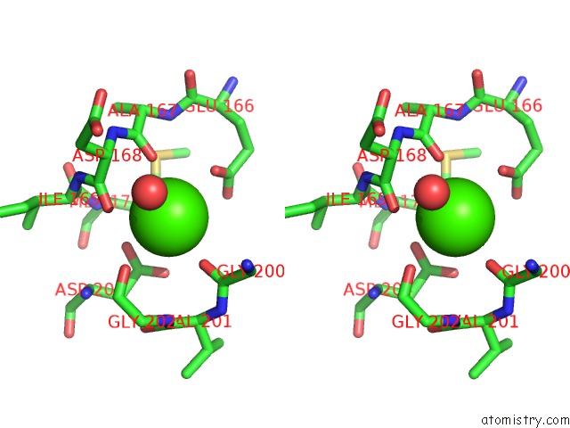
Stereo pair view
Mono view
Stereo pair view
A full contact list of Calcium with other atoms in the Ca binding
site number 2 of Gelatinase A (Full-Length) within 5.0Å range:
|
Calcium binding site 3 out of 3 in 1ck7
Go back to
Calcium binding site 3 out
of 3 in the Gelatinase A (Full-Length)
Mono view
Stereo pair view
Mono view
Stereo pair view
A full contact list of Calcium with other atoms in the Ca binding
site number 3 of Gelatinase A (Full-Length) within 5.0Å range:
|
Reference:
E.Morgunova,
A.Tuuttila,
U.Bergmann,
M.Isupov,
Y.Lindqvist,
G.Schneider,
K.Tryggvason.
Structure of Human Pro-Matrix Metalloproteinase-2: Activation Mechanism Revealed. Science V. 284 1667 1999.
ISSN: ISSN 0036-8075
PubMed: 10356396
DOI: 10.1126/SCIENCE.284.5420.1667
Page generated: Mon Jul 7 14:02:40 2025
ISSN: ISSN 0036-8075
PubMed: 10356396
DOI: 10.1126/SCIENCE.284.5420.1667
Last articles
F in 4IF4F in 4IIZ
F in 4IDQ
F in 4IGH
F in 4IGA
F in 4IBJ
F in 4IFY
F in 4IFV
F in 4IDO
F in 4ICC