Calcium »
PDB 1cxk-1dds »
1d2s »
Calcium in PDB 1d2s: Crystal Structure of the N-Terminal Laminin G-Like Domain of Shbg in Complex with Dihydrotestosterone
Protein crystallography data
The structure of Crystal Structure of the N-Terminal Laminin G-Like Domain of Shbg in Complex with Dihydrotestosterone, PDB code: 1d2s
was solved by
I.Grishkovskaya,
G.V.Avvakumov,
G.Sklenar,
D.Dales,
G.L.Hammond,
Y.A.Muller,
with X-Ray Crystallography technique. A brief refinement statistics is given in the table below:
| Resolution Low / High (Å) | 40.00 / 1.55 |
| Space group | H 3 2 |
| Cell size a, b, c (Å), α, β, γ (°) | 104.040, 104.040, 84.430, 90.00, 90.00, 120.00 |
| R / Rfree (%) | 20.5 / 25.1 |
Calcium Binding Sites:
The binding sites of Calcium atom in the Crystal Structure of the N-Terminal Laminin G-Like Domain of Shbg in Complex with Dihydrotestosterone
(pdb code 1d2s). This binding sites where shown within
5.0 Angstroms radius around Calcium atom.
In total 2 binding sites of Calcium where determined in the Crystal Structure of the N-Terminal Laminin G-Like Domain of Shbg in Complex with Dihydrotestosterone, PDB code: 1d2s:
Jump to Calcium binding site number: 1; 2;
In total 2 binding sites of Calcium where determined in the Crystal Structure of the N-Terminal Laminin G-Like Domain of Shbg in Complex with Dihydrotestosterone, PDB code: 1d2s:
Jump to Calcium binding site number: 1; 2;
Calcium binding site 1 out of 2 in 1d2s
Go back to
Calcium binding site 1 out
of 2 in the Crystal Structure of the N-Terminal Laminin G-Like Domain of Shbg in Complex with Dihydrotestosterone
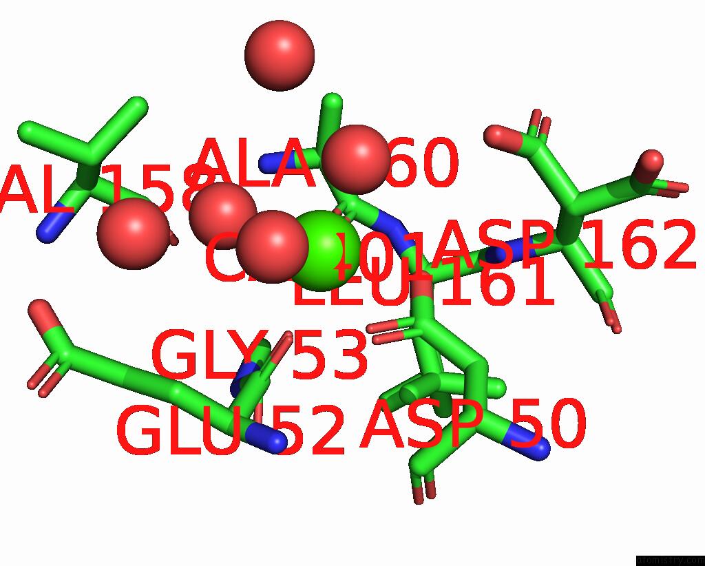
Mono view
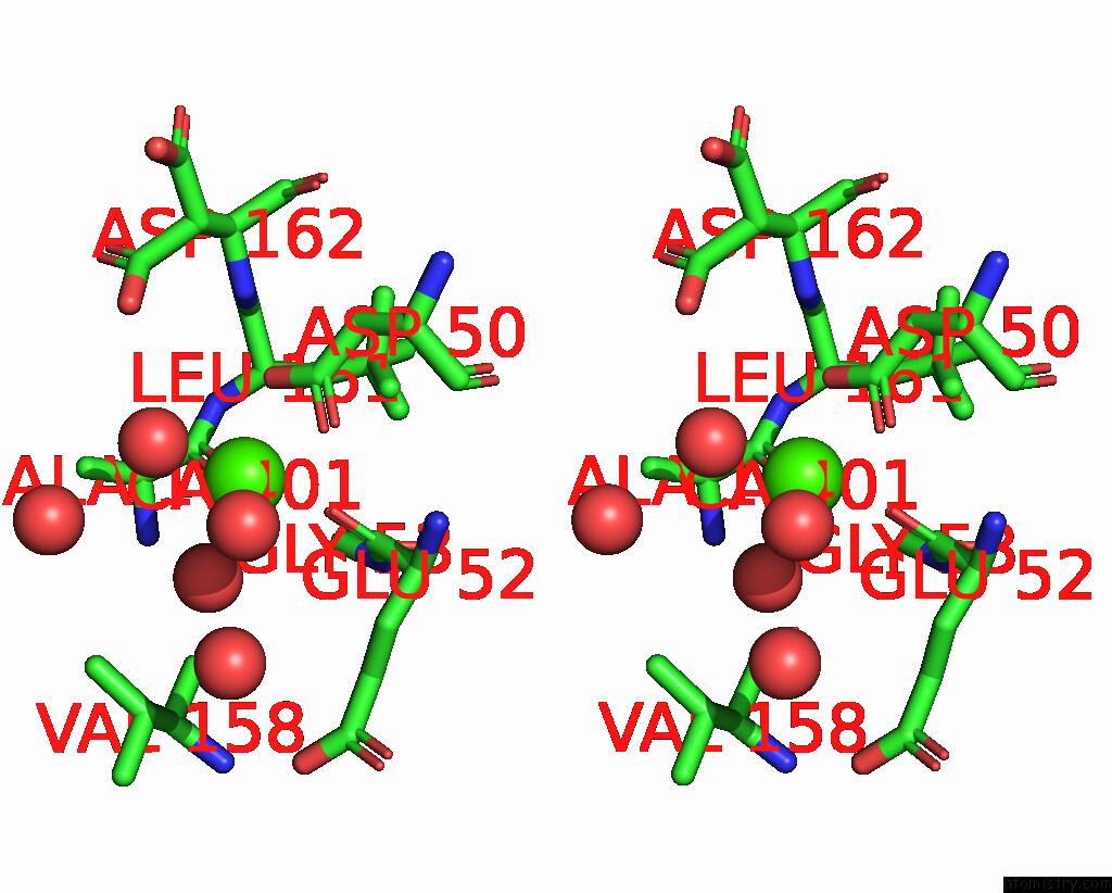
Stereo pair view
Mono view
Stereo pair view
A full contact list of Calcium with other atoms in the Ca binding
site number 1 of Crystal Structure of the N-Terminal Laminin G-Like Domain of Shbg in Complex with Dihydrotestosterone within 5.0Å range:
|
Calcium binding site 2 out of 2 in 1d2s
Go back to
Calcium binding site 2 out
of 2 in the Crystal Structure of the N-Terminal Laminin G-Like Domain of Shbg in Complex with Dihydrotestosterone
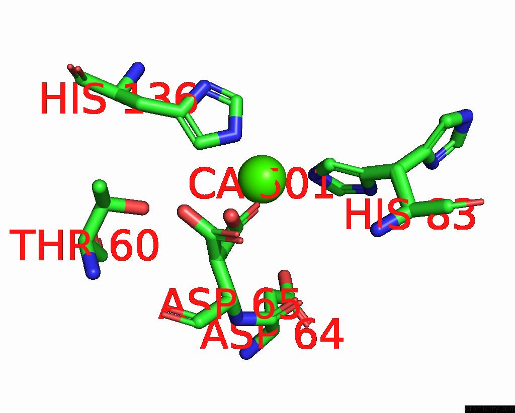
Mono view
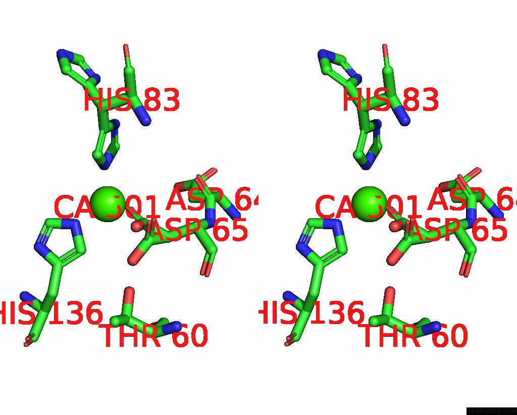
Stereo pair view
Mono view
Stereo pair view
A full contact list of Calcium with other atoms in the Ca binding
site number 2 of Crystal Structure of the N-Terminal Laminin G-Like Domain of Shbg in Complex with Dihydrotestosterone within 5.0Å range:
|
Reference:
I.Grishkovskaya,
G.V.Avvakumov,
G.Sklenar,
D.Dales,
G.L.Hammond,
Y.A.Muller.
Crystal Structure of Human Sex Hormone-Binding Globulin: Steroid Transport By A Laminin G-Like Domain. Embo J. V. 19 504 2000.
ISSN: ISSN 0261-4189
PubMed: 10675319
DOI: 10.1093/EMBOJ/19.4.504
Page generated: Thu Jul 11 07:22:59 2024
ISSN: ISSN 0261-4189
PubMed: 10675319
DOI: 10.1093/EMBOJ/19.4.504
Last articles
Zn in 9J0NZn in 9J0O
Zn in 9J0P
Zn in 9FJX
Zn in 9EKB
Zn in 9C0F
Zn in 9CAH
Zn in 9CH0
Zn in 9CH3
Zn in 9CH1