Calcium »
PDB 1cxk-1dds »
1d2v »
Calcium in PDB 1d2v: Crystal Structure of Bromide-Bound Human Myeloperoxidase Isoform C at pH 5.5
Enzymatic activity of Crystal Structure of Bromide-Bound Human Myeloperoxidase Isoform C at pH 5.5
All present enzymatic activity of Crystal Structure of Bromide-Bound Human Myeloperoxidase Isoform C at pH 5.5:
1.11.1.7;
1.11.1.7;
Protein crystallography data
The structure of Crystal Structure of Bromide-Bound Human Myeloperoxidase Isoform C at pH 5.5, PDB code: 1d2v
was solved by
T.J.Fiedler,
C.A.Davey,
R.E.Fenna,
with X-Ray Crystallography technique. A brief refinement statistics is given in the table below:
| Resolution Low / High (Å) | 30.00 / 1.75 |
| Space group | P 1 21 1 |
| Cell size a, b, c (Å), α, β, γ (°) | 111.155, 63.488, 92.476, 90.00, 97.36, 90.00 |
| R / Rfree (%) | 24.3 / 29.6 |
Other elements in 1d2v:
The structure of Crystal Structure of Bromide-Bound Human Myeloperoxidase Isoform C at pH 5.5 also contains other interesting chemical elements:
| Bromine | (Br) | 8 atoms |
| Iron | (Fe) | 2 atoms |
Calcium Binding Sites:
The binding sites of Calcium atom in the Crystal Structure of Bromide-Bound Human Myeloperoxidase Isoform C at pH 5.5
(pdb code 1d2v). This binding sites where shown within
5.0 Angstroms radius around Calcium atom.
In total 2 binding sites of Calcium where determined in the Crystal Structure of Bromide-Bound Human Myeloperoxidase Isoform C at pH 5.5, PDB code: 1d2v:
Jump to Calcium binding site number: 1; 2;
In total 2 binding sites of Calcium where determined in the Crystal Structure of Bromide-Bound Human Myeloperoxidase Isoform C at pH 5.5, PDB code: 1d2v:
Jump to Calcium binding site number: 1; 2;
Calcium binding site 1 out of 2 in 1d2v
Go back to
Calcium binding site 1 out
of 2 in the Crystal Structure of Bromide-Bound Human Myeloperoxidase Isoform C at pH 5.5
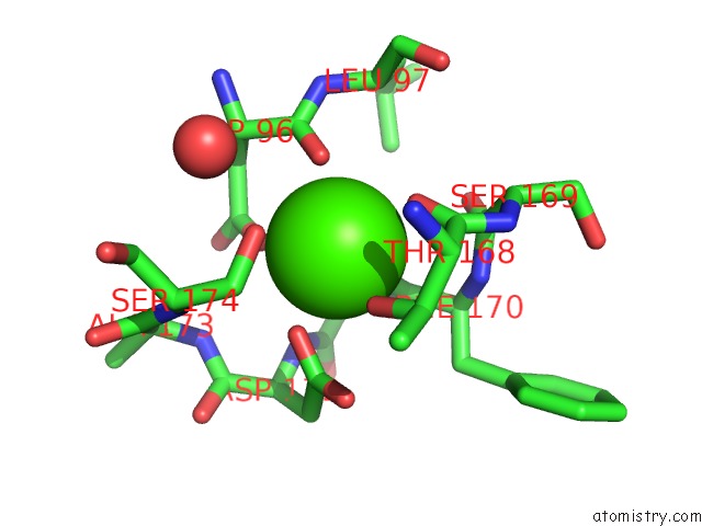
Mono view
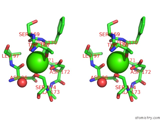
Stereo pair view
Mono view
Stereo pair view
A full contact list of Calcium with other atoms in the Ca binding
site number 1 of Crystal Structure of Bromide-Bound Human Myeloperoxidase Isoform C at pH 5.5 within 5.0Å range:
|
Calcium binding site 2 out of 2 in 1d2v
Go back to
Calcium binding site 2 out
of 2 in the Crystal Structure of Bromide-Bound Human Myeloperoxidase Isoform C at pH 5.5
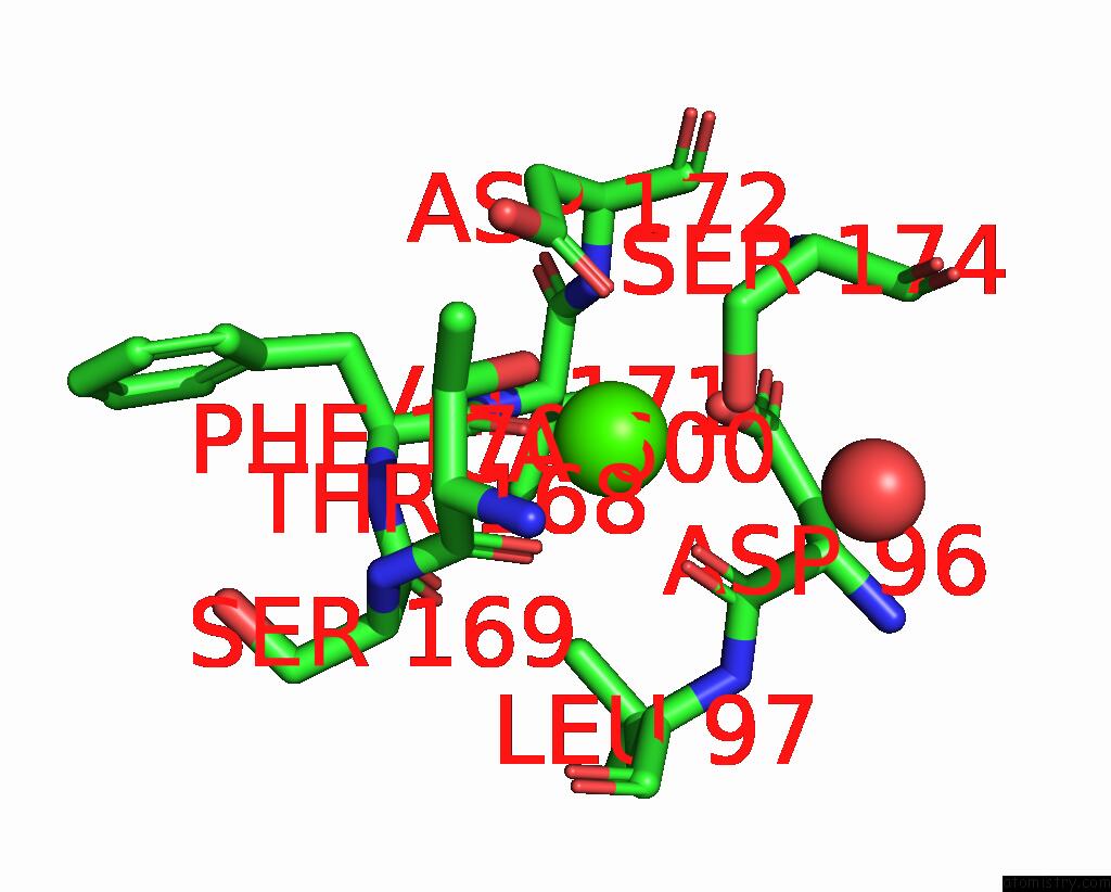
Mono view
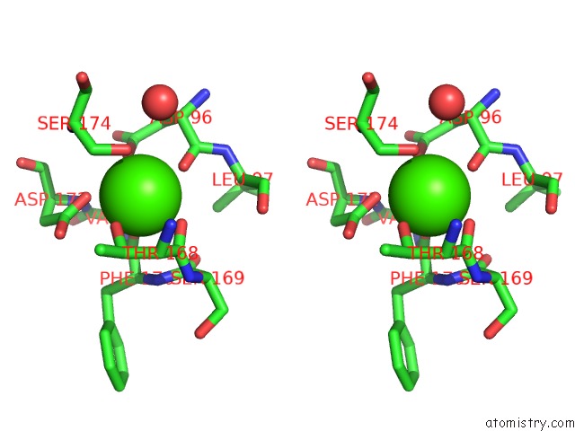
Stereo pair view
Mono view
Stereo pair view
A full contact list of Calcium with other atoms in the Ca binding
site number 2 of Crystal Structure of Bromide-Bound Human Myeloperoxidase Isoform C at pH 5.5 within 5.0Å range:
|
Reference:
T.J.Fiedler,
C.A.Davey,
R.E.Fenna.
X-Ray Crystal Structure and Characterization of Halide-Binding Sites of Human Myeloperoxidase at 1.8 A Resolution. J.Biol.Chem. V. 275 11964 2000.
ISSN: ISSN 0021-9258
PubMed: 10766826
DOI: 10.1074/JBC.275.16.11964
Page generated: Thu Jul 11 07:23:01 2024
ISSN: ISSN 0021-9258
PubMed: 10766826
DOI: 10.1074/JBC.275.16.11964
Last articles
Zn in 9MJ5Zn in 9HNW
Zn in 9G0L
Zn in 9FNE
Zn in 9DZN
Zn in 9E0I
Zn in 9D32
Zn in 9DAK
Zn in 8ZXC
Zn in 8ZUF