Calcium »
PDB 1cxk-1dds »
1ddr »
Calcium in PDB 1ddr: Molecule: Dihydrofolate Reductase (E.C.1.5.1.3) Complexed with Methotrexate and Urea
Enzymatic activity of Molecule: Dihydrofolate Reductase (E.C.1.5.1.3) Complexed with Methotrexate and Urea
All present enzymatic activity of Molecule: Dihydrofolate Reductase (E.C.1.5.1.3) Complexed with Methotrexate and Urea:
1.5.1.3;
1.5.1.3;
Protein crystallography data
The structure of Molecule: Dihydrofolate Reductase (E.C.1.5.1.3) Complexed with Methotrexate and Urea, PDB code: 1ddr
was solved by
H.P.Yennawar,
G.K.Farber,
with X-Ray Crystallography technique. A brief refinement statistics is given in the table below:
| Resolution Low / High (Å) | 10.00 / 2.45 |
| Space group | P 61 |
| Cell size a, b, c (Å), α, β, γ (°) | 93.181, 93.185, 73.886, 90.00, 90.00, 120.00 |
| R / Rfree (%) | 15.6 / 17.1 |
Other elements in 1ddr:
The structure of Molecule: Dihydrofolate Reductase (E.C.1.5.1.3) Complexed with Methotrexate and Urea also contains other interesting chemical elements:
| Chlorine | (Cl) | 2 atoms |
Calcium Binding Sites:
The binding sites of Calcium atom in the Molecule: Dihydrofolate Reductase (E.C.1.5.1.3) Complexed with Methotrexate and Urea
(pdb code 1ddr). This binding sites where shown within
5.0 Angstroms radius around Calcium atom.
In total only one binding site of Calcium was determined in the Molecule: Dihydrofolate Reductase (E.C.1.5.1.3) Complexed with Methotrexate and Urea, PDB code: 1ddr:
In total only one binding site of Calcium was determined in the Molecule: Dihydrofolate Reductase (E.C.1.5.1.3) Complexed with Methotrexate and Urea, PDB code: 1ddr:
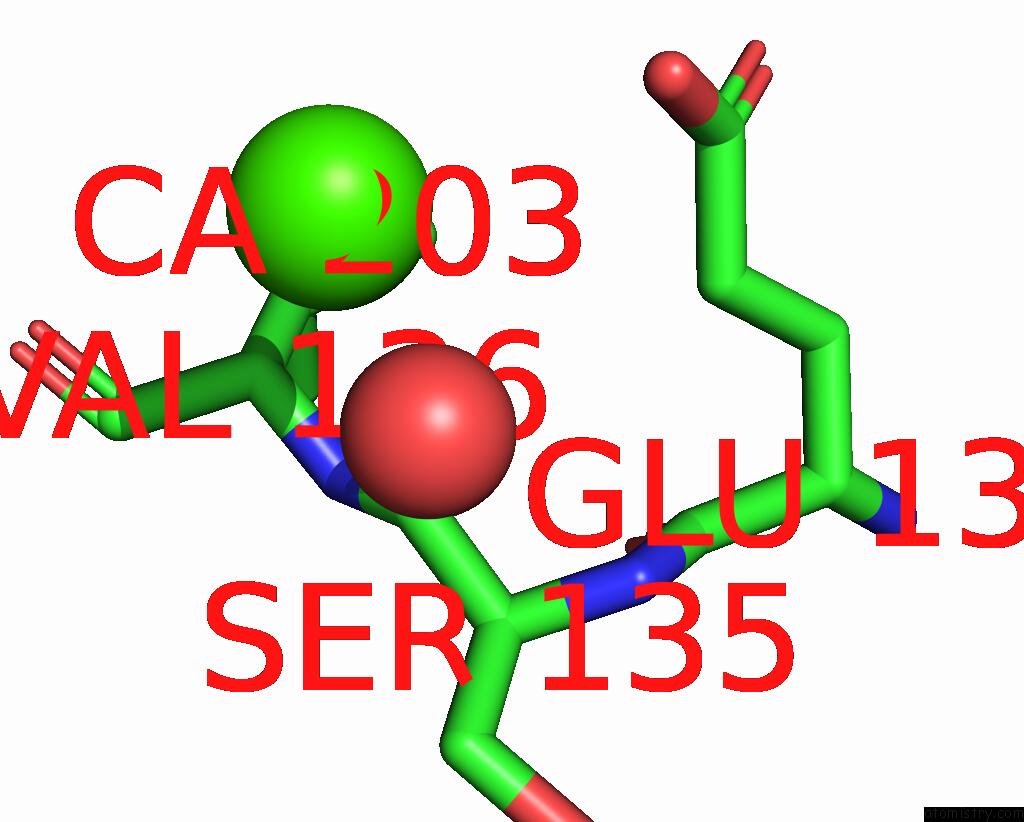
Calcium binding site 1 out of 1 in 1ddr
Go back to
Calcium binding site 1 out
of 1 in the Molecule: Dihydrofolate Reductase (E.C.1.5.1.3) Complexed with Methotrexate and Urea
Mono view

Stereo pair view
Mono view
Stereo pair view
A full contact list of Calcium with other atoms in the Ca binding
site number 1 of Molecule: Dihydrofolate Reductase (E.C.1.5.1.3) Complexed with Methotrexate and Urea within 5.0Å range:
|
Reference:
J.Dunbar,
H.P.Yennawar,
S.Banerjee,
J.Luo,
G.K.Farber.
The Effect of Denaturants on Protein Structure. Protein Sci. V. 6 1727 1997.
ISSN: ISSN 0961-8368
PubMed: 9260285
Page generated: Thu Jul 11 07:30:57 2024
ISSN: ISSN 0961-8368
PubMed: 9260285
Last articles
Zn in 9MJ5Zn in 9HNW
Zn in 9G0L
Zn in 9FNE
Zn in 9DZN
Zn in 9E0I
Zn in 9D32
Zn in 9DAK
Zn in 8ZXC
Zn in 8ZUF