Calcium »
PDB 1de4-1dv8 »
1dl2 »
Calcium in PDB 1dl2: Crystal Structure of Class I Alpha-1,2-Mannosidase From Saccharomyces Cerevisiae at 1.54 Angstrom Resolution
Enzymatic activity of Crystal Structure of Class I Alpha-1,2-Mannosidase From Saccharomyces Cerevisiae at 1.54 Angstrom Resolution
All present enzymatic activity of Crystal Structure of Class I Alpha-1,2-Mannosidase From Saccharomyces Cerevisiae at 1.54 Angstrom Resolution:
3.2.1.113;
3.2.1.113;
Protein crystallography data
The structure of Crystal Structure of Class I Alpha-1,2-Mannosidase From Saccharomyces Cerevisiae at 1.54 Angstrom Resolution, PDB code: 1dl2
was solved by
F.Vallee,
F.Lipari,
P.Yip,
A.Herscovics,
P.L.Howell,
with X-Ray Crystallography technique. A brief refinement statistics is given in the table below:
| Resolution Low / High (Å) | 50.00 / 1.54 |
| Space group | P 31 2 1 |
| Cell size a, b, c (Å), α, β, γ (°) | 88.397, 88.397, 153.220, 90.00, 90.00, 120.00 |
| R / Rfree (%) | 20.9 / 22.7 |
Calcium Binding Sites:
The binding sites of Calcium atom in the Crystal Structure of Class I Alpha-1,2-Mannosidase From Saccharomyces Cerevisiae at 1.54 Angstrom Resolution
(pdb code 1dl2). This binding sites where shown within
5.0 Angstroms radius around Calcium atom.
In total only one binding site of Calcium was determined in the Crystal Structure of Class I Alpha-1,2-Mannosidase From Saccharomyces Cerevisiae at 1.54 Angstrom Resolution, PDB code: 1dl2:
In total only one binding site of Calcium was determined in the Crystal Structure of Class I Alpha-1,2-Mannosidase From Saccharomyces Cerevisiae at 1.54 Angstrom Resolution, PDB code: 1dl2:
Calcium binding site 1 out of 1 in 1dl2
Go back to
Calcium binding site 1 out
of 1 in the Crystal Structure of Class I Alpha-1,2-Mannosidase From Saccharomyces Cerevisiae at 1.54 Angstrom Resolution
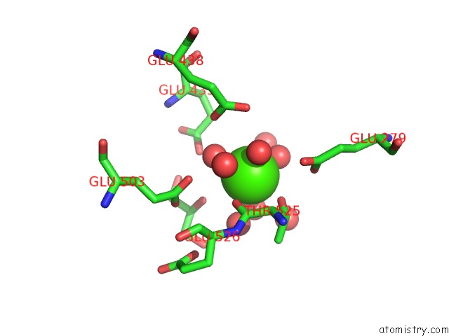
Mono view
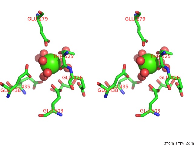
Stereo pair view
Mono view
Stereo pair view
A full contact list of Calcium with other atoms in the Ca binding
site number 1 of Crystal Structure of Class I Alpha-1,2-Mannosidase From Saccharomyces Cerevisiae at 1.54 Angstrom Resolution within 5.0Å range:
|
Reference:
F.Vallee,
F.Lipari,
P.Yip,
B.Sleno,
A.Herscovics,
P.L.Howell.
Crystal Structure of A Class I ALPHA1,2-Mannosidase Involved in N-Glycan Processing and Endoplasmic Reticulum Quality Control. Embo J. V. 19 581 2000.
ISSN: ISSN 0261-4189
PubMed: 10675327
DOI: 10.1093/EMBOJ/19.4.581
Page generated: Thu Jul 11 07:35:22 2024
ISSN: ISSN 0261-4189
PubMed: 10675327
DOI: 10.1093/EMBOJ/19.4.581
Last articles
Zn in 9MJ5Zn in 9HNW
Zn in 9G0L
Zn in 9FNE
Zn in 9DZN
Zn in 9E0I
Zn in 9D32
Zn in 9DAK
Zn in 8ZXC
Zn in 8ZUF