Calcium »
PDB 1de4-1dv8 »
1dmu »
Calcium in PDB 1dmu: Crystal Structure of the Restriction Endonuclease Bgli (E.C.3.1.21.4) Bound to Its Dna Recognition Sequence
Enzymatic activity of Crystal Structure of the Restriction Endonuclease Bgli (E.C.3.1.21.4) Bound to Its Dna Recognition Sequence
All present enzymatic activity of Crystal Structure of the Restriction Endonuclease Bgli (E.C.3.1.21.4) Bound to Its Dna Recognition Sequence:
3.1.21.4;
3.1.21.4;
Protein crystallography data
The structure of Crystal Structure of the Restriction Endonuclease Bgli (E.C.3.1.21.4) Bound to Its Dna Recognition Sequence, PDB code: 1dmu
was solved by
M.Newman,
K.Lunnen,
G.Wilson,
J.Greci,
I.Schildkraut,
S.E.V.Phillips,
with X-Ray Crystallography technique. A brief refinement statistics is given in the table below:
| Resolution Low / High (Å) | 10.00 / 2.20 |
| Space group | C 2 2 21 |
| Cell size a, b, c (Å), α, β, γ (°) | 78.480, 81.600, 117.060, 90.00, 90.00, 90.00 |
| R / Rfree (%) | 17.7 / 23.9 |
Calcium Binding Sites:
The binding sites of Calcium atom in the Crystal Structure of the Restriction Endonuclease Bgli (E.C.3.1.21.4) Bound to Its Dna Recognition Sequence
(pdb code 1dmu). This binding sites where shown within
5.0 Angstroms radius around Calcium atom.
In total 4 binding sites of Calcium where determined in the Crystal Structure of the Restriction Endonuclease Bgli (E.C.3.1.21.4) Bound to Its Dna Recognition Sequence, PDB code: 1dmu:
Jump to Calcium binding site number: 1; 2; 3; 4;
In total 4 binding sites of Calcium where determined in the Crystal Structure of the Restriction Endonuclease Bgli (E.C.3.1.21.4) Bound to Its Dna Recognition Sequence, PDB code: 1dmu:
Jump to Calcium binding site number: 1; 2; 3; 4;
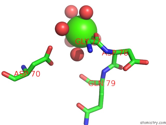
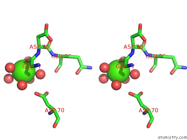
Calcium binding site 1 out of 4 in 1dmu
Go back to
Calcium binding site 1 out
of 4 in the Crystal Structure of the Restriction Endonuclease Bgli (E.C.3.1.21.4) Bound to Its Dna Recognition Sequence
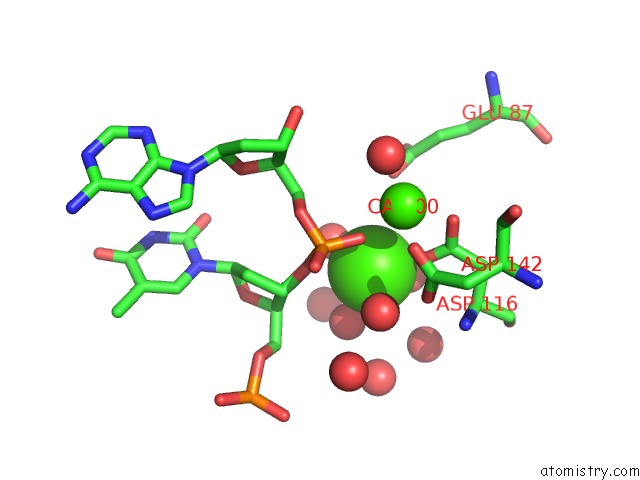
Mono view
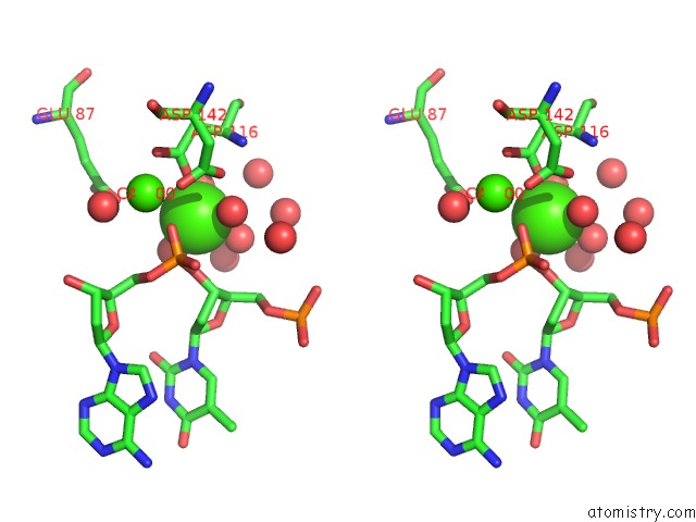
Stereo pair view
Mono view
Stereo pair view
A full contact list of Calcium with other atoms in the Ca binding
site number 1 of Crystal Structure of the Restriction Endonuclease Bgli (E.C.3.1.21.4) Bound to Its Dna Recognition Sequence within 5.0Å range:
|
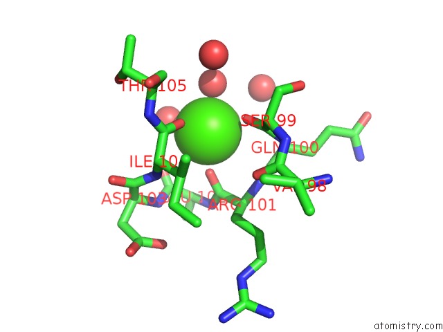
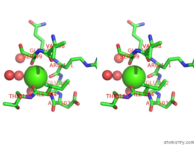
Calcium binding site 2 out of 4 in 1dmu
Go back to
Calcium binding site 2 out
of 4 in the Crystal Structure of the Restriction Endonuclease Bgli (E.C.3.1.21.4) Bound to Its Dna Recognition Sequence
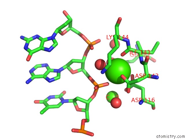
Mono view
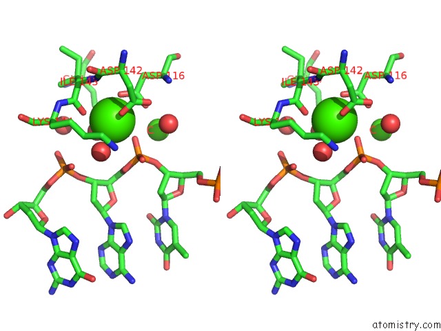
Stereo pair view
Mono view
Stereo pair view
A full contact list of Calcium with other atoms in the Ca binding
site number 2 of Crystal Structure of the Restriction Endonuclease Bgli (E.C.3.1.21.4) Bound to Its Dna Recognition Sequence within 5.0Å range:
|
Calcium binding site 3 out of 4 in 1dmu
Go back to
Calcium binding site 3 out
of 4 in the Crystal Structure of the Restriction Endonuclease Bgli (E.C.3.1.21.4) Bound to Its Dna Recognition Sequence
Mono view
Stereo pair view
Mono view
Stereo pair view
A full contact list of Calcium with other atoms in the Ca binding
site number 3 of Crystal Structure of the Restriction Endonuclease Bgli (E.C.3.1.21.4) Bound to Its Dna Recognition Sequence within 5.0Å range:
|
Calcium binding site 4 out of 4 in 1dmu
Go back to
Calcium binding site 4 out
of 4 in the Crystal Structure of the Restriction Endonuclease Bgli (E.C.3.1.21.4) Bound to Its Dna Recognition Sequence
Mono view
Stereo pair view
Mono view
Stereo pair view
A full contact list of Calcium with other atoms in the Ca binding
site number 4 of Crystal Structure of the Restriction Endonuclease Bgli (E.C.3.1.21.4) Bound to Its Dna Recognition Sequence within 5.0Å range:
|
Reference:
M.Newman,
K.Lunnen,
G.Wilson,
J.Greci,
I.Schildkraut,
S.E.Phillips.
Crystal Structure of Restriction Endonuclease Bgli Bound to Its Interrupted Dna Recognition Sequence. Embo J. V. 17 5466 1998.
ISSN: ISSN 0261-4189
PubMed: 9736624
DOI: 10.1093/EMBOJ/17.18.5466
Page generated: Thu Jul 11 07:35:49 2024
ISSN: ISSN 0261-4189
PubMed: 9736624
DOI: 10.1093/EMBOJ/17.18.5466
Last articles
Zn in 9MJ5Zn in 9HNW
Zn in 9G0L
Zn in 9FNE
Zn in 9DZN
Zn in 9E0I
Zn in 9D32
Zn in 9DAK
Zn in 8ZXC
Zn in 8ZUF