Calcium »
PDB 1de4-1dv8 »
1dr3 »
Calcium in PDB 1dr3: 2.3 Angstroms Crystal Structure of Chicken Liver Dihydrofolate Reductase Complexed with Thionadp+ and Biopterin
Enzymatic activity of 2.3 Angstroms Crystal Structure of Chicken Liver Dihydrofolate Reductase Complexed with Thionadp+ and Biopterin
All present enzymatic activity of 2.3 Angstroms Crystal Structure of Chicken Liver Dihydrofolate Reductase Complexed with Thionadp+ and Biopterin:
1.5.1.3;
1.5.1.3;
Protein crystallography data
The structure of 2.3 Angstroms Crystal Structure of Chicken Liver Dihydrofolate Reductase Complexed with Thionadp+ and Biopterin, PDB code: 1dr3
was solved by
M.A.Mctigue,
J.F.Davies /Ii,
B.T.Kaufman,
N.-H.Xuong,
J.Kraut,
with X-Ray Crystallography technique. A brief refinement statistics is given in the table below:
| Resolution Low / High (Å) | N/A / 2.30 |
| Space group | C 1 2 1 |
| Cell size a, b, c (Å), α, β, γ (°) | 89.200, 48.310, 64.230, 90.00, 124.80, 90.00 |
| R / Rfree (%) | n/a / n/a |
Calcium Binding Sites:
The binding sites of Calcium atom in the 2.3 Angstroms Crystal Structure of Chicken Liver Dihydrofolate Reductase Complexed with Thionadp+ and Biopterin
(pdb code 1dr3). This binding sites where shown within
5.0 Angstroms radius around Calcium atom.
In total only one binding site of Calcium was determined in the 2.3 Angstroms Crystal Structure of Chicken Liver Dihydrofolate Reductase Complexed with Thionadp+ and Biopterin, PDB code: 1dr3:
In total only one binding site of Calcium was determined in the 2.3 Angstroms Crystal Structure of Chicken Liver Dihydrofolate Reductase Complexed with Thionadp+ and Biopterin, PDB code: 1dr3:
Calcium binding site 1 out of 1 in 1dr3
Go back to
Calcium binding site 1 out
of 1 in the 2.3 Angstroms Crystal Structure of Chicken Liver Dihydrofolate Reductase Complexed with Thionadp+ and Biopterin
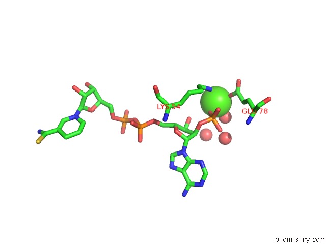
Mono view
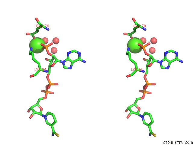
Stereo pair view
Mono view
Stereo pair view
A full contact list of Calcium with other atoms in the Ca binding
site number 1 of 2.3 Angstroms Crystal Structure of Chicken Liver Dihydrofolate Reductase Complexed with Thionadp+ and Biopterin within 5.0Å range:
|
Reference:
M.A.Mctigue,
J.F.Davies 2Nd.,
B.T.Kaufman,
J.Kraut.
Crystal Structures of Chicken Liver Dihydrofolate Reductase: Binary Thionadp+ and Ternary Thionadp+.Biopterin Complexes. Biochemistry V. 32 6855 1993.
ISSN: ISSN 0006-2960
PubMed: 8334118
DOI: 10.1021/BI00078A008
Page generated: Thu Jul 11 07:37:56 2024
ISSN: ISSN 0006-2960
PubMed: 8334118
DOI: 10.1021/BI00078A008
Last articles
Zn in 9MJ5Zn in 9HNW
Zn in 9G0L
Zn in 9FNE
Zn in 9DZN
Zn in 9E0I
Zn in 9D32
Zn in 9DAK
Zn in 8ZXC
Zn in 8ZUF