Calcium »
PDB 1de4-1dv8 »
1dr6 »
Calcium in PDB 1dr6: Crystal Structures of Organomercurial-Activated Chicken Liver Dihydrofolate Reductase Complexes
Enzymatic activity of Crystal Structures of Organomercurial-Activated Chicken Liver Dihydrofolate Reductase Complexes
All present enzymatic activity of Crystal Structures of Organomercurial-Activated Chicken Liver Dihydrofolate Reductase Complexes:
1.5.1.3;
1.5.1.3;
Protein crystallography data
The structure of Crystal Structures of Organomercurial-Activated Chicken Liver Dihydrofolate Reductase Complexes, PDB code: 1dr6
was solved by
M.A.Mctigue,
J.F.Davies /Ii,
B.T.Kaufman,
N.-H.Xuong,
J.Kraut,
with X-Ray Crystallography technique. A brief refinement statistics is given in the table below:
| Resolution Low / High (Å) | N/A / 2.40 |
| Space group | C 1 2 1 |
| Cell size a, b, c (Å), α, β, γ (°) | 88.720, 48.740, 63.870, 90.00, 124.70, 90.00 |
| R / Rfree (%) | n/a / n/a |
Other elements in 1dr6:
The structure of Crystal Structures of Organomercurial-Activated Chicken Liver Dihydrofolate Reductase Complexes also contains other interesting chemical elements:
| Mercury | (Hg) | 2 atoms |
Calcium Binding Sites:
The binding sites of Calcium atom in the Crystal Structures of Organomercurial-Activated Chicken Liver Dihydrofolate Reductase Complexes
(pdb code 1dr6). This binding sites where shown within
5.0 Angstroms radius around Calcium atom.
In total only one binding site of Calcium was determined in the Crystal Structures of Organomercurial-Activated Chicken Liver Dihydrofolate Reductase Complexes, PDB code: 1dr6:
In total only one binding site of Calcium was determined in the Crystal Structures of Organomercurial-Activated Chicken Liver Dihydrofolate Reductase Complexes, PDB code: 1dr6:
Calcium binding site 1 out of 1 in 1dr6
Go back to
Calcium binding site 1 out
of 1 in the Crystal Structures of Organomercurial-Activated Chicken Liver Dihydrofolate Reductase Complexes
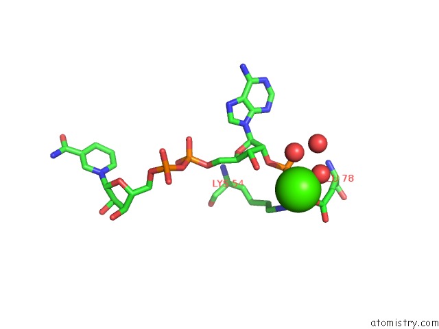
Mono view
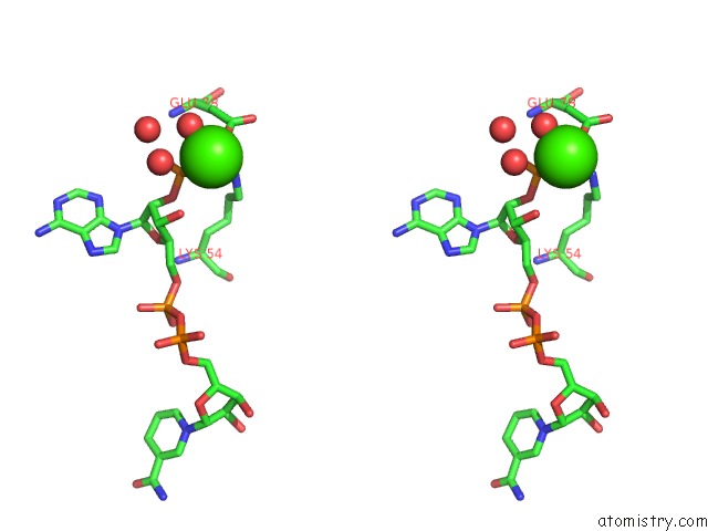
Stereo pair view
Mono view
Stereo pair view
A full contact list of Calcium with other atoms in the Ca binding
site number 1 of Crystal Structures of Organomercurial-Activated Chicken Liver Dihydrofolate Reductase Complexes within 5.0Å range:
|
Reference:
M.A.Mctigue,
J.F.Davies Ii,
B.T.Kaufman,
N.-H.Xuong,
J.Kraut.
Crystal Structures of Organomercurial-Activated Chicken Liver Dihydrofolate Reductase Complexes To Be Published.
Page generated: Mon Jul 7 14:28:59 2025
Last articles
Cl in 5RAECl in 5RAD
Cl in 5RAC
Cl in 5RAB
Cl in 5RA9
Cl in 5RAA
Cl in 5RA8
Cl in 5RA7
Cl in 5RA4
Cl in 5RA6