Calcium »
PDB 1dva-1edh »
1dva »
Calcium in PDB 1dva: Crystal Structure of the Complex Between the Peptide Exosite Inhibitor E-76 and Coagulation Factor Viia
Enzymatic activity of Crystal Structure of the Complex Between the Peptide Exosite Inhibitor E-76 and Coagulation Factor Viia
All present enzymatic activity of Crystal Structure of the Complex Between the Peptide Exosite Inhibitor E-76 and Coagulation Factor Viia:
3.4.21.21;
3.4.21.21;
Protein crystallography data
The structure of Crystal Structure of the Complex Between the Peptide Exosite Inhibitor E-76 and Coagulation Factor Viia, PDB code: 1dva
was solved by
C.Eigenbrot,
M.H.Ultsch,
with X-Ray Crystallography technique. A brief refinement statistics is given in the table below:
| Resolution Low / High (Å) | 50.00 / 3.00 |
| Space group | P 1 21 1 |
| Cell size a, b, c (Å), α, β, γ (°) | 70.490, 55.260, 111.730, 90.00, 99.48, 90.00 |
| R / Rfree (%) | 22.5 / 29.5 |
Other elements in 1dva:
The structure of Crystal Structure of the Complex Between the Peptide Exosite Inhibitor E-76 and Coagulation Factor Viia also contains other interesting chemical elements:
| Arsenic | (As) | 2 atoms |
Calcium Binding Sites:
The binding sites of Calcium atom in the Crystal Structure of the Complex Between the Peptide Exosite Inhibitor E-76 and Coagulation Factor Viia
(pdb code 1dva). This binding sites where shown within
5.0 Angstroms radius around Calcium atom.
In total 4 binding sites of Calcium where determined in the Crystal Structure of the Complex Between the Peptide Exosite Inhibitor E-76 and Coagulation Factor Viia, PDB code: 1dva:
Jump to Calcium binding site number: 1; 2; 3; 4;
In total 4 binding sites of Calcium where determined in the Crystal Structure of the Complex Between the Peptide Exosite Inhibitor E-76 and Coagulation Factor Viia, PDB code: 1dva:
Jump to Calcium binding site number: 1; 2; 3; 4;
Calcium binding site 1 out of 4 in 1dva
Go back to
Calcium binding site 1 out
of 4 in the Crystal Structure of the Complex Between the Peptide Exosite Inhibitor E-76 and Coagulation Factor Viia

Mono view
Stereo pair view
Mono view
Stereo pair view
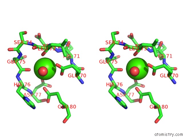
A full contact list of Calcium with other atoms in the Ca binding
site number 1 of Crystal Structure of the Complex Between the Peptide Exosite Inhibitor E-76 and Coagulation Factor Viia within 5.0Å range:
|
Calcium binding site 2 out of 4 in 1dva
Go back to
Calcium binding site 2 out
of 4 in the Crystal Structure of the Complex Between the Peptide Exosite Inhibitor E-76 and Coagulation Factor Viia

Mono view

Stereo pair view
Mono view
Stereo pair view
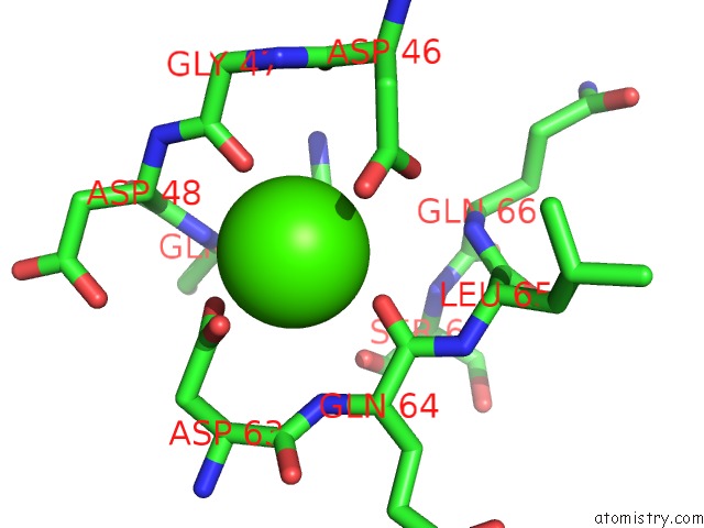
A full contact list of Calcium with other atoms in the Ca binding
site number 2 of Crystal Structure of the Complex Between the Peptide Exosite Inhibitor E-76 and Coagulation Factor Viia within 5.0Å range:
|
Calcium binding site 3 out of 4 in 1dva
Go back to
Calcium binding site 3 out
of 4 in the Crystal Structure of the Complex Between the Peptide Exosite Inhibitor E-76 and Coagulation Factor Viia

Mono view

Stereo pair view
Mono view
Stereo pair view
A full contact list of Calcium with other atoms in the Ca binding
site number 3 of Crystal Structure of the Complex Between the Peptide Exosite Inhibitor E-76 and Coagulation Factor Viia within 5.0Å range:
|
Calcium binding site 4 out of 4 in 1dva
Go back to
Calcium binding site 4 out
of 4 in the Crystal Structure of the Complex Between the Peptide Exosite Inhibitor E-76 and Coagulation Factor Viia
Mono view

Stereo pair view
Mono view
Stereo pair view
A full contact list of Calcium with other atoms in the Ca binding
site number 4 of Crystal Structure of the Complex Between the Peptide Exosite Inhibitor E-76 and Coagulation Factor Viia within 5.0Å range:
|
Reference:
M.S.Dennis,
C.Eigenbrot,
N.J.Skelton,
M.H.Ultsch,
L.Santell,
M.A.Dwyer,
M.P.O'connell,
R.A.Lazarus.
Peptide Exosite Inhibitors of Factor Viia As Anticoagulants. Nature V. 404 465 2000.
ISSN: ISSN 0028-0836
PubMed: 10761907
DOI: 10.1038/35006574
Page generated: Thu Jul 11 07:41:22 2024
ISSN: ISSN 0028-0836
PubMed: 10761907
DOI: 10.1038/35006574
Last articles
Zn in 9J0NZn in 9J0O
Zn in 9J0P
Zn in 9FJX
Zn in 9EKB
Zn in 9C0F
Zn in 9CAH
Zn in 9CH0
Zn in 9CH3
Zn in 9CH1