Calcium »
PDB 1dva-1edh »
1e8t »
Calcium in PDB 1e8t: Structure of the Multifunctional Paramyxovirus Hemagglutinin- Neuraminidase
Enzymatic activity of Structure of the Multifunctional Paramyxovirus Hemagglutinin- Neuraminidase
All present enzymatic activity of Structure of the Multifunctional Paramyxovirus Hemagglutinin- Neuraminidase:
3.2.1.18;
3.2.1.18;
Protein crystallography data
The structure of Structure of the Multifunctional Paramyxovirus Hemagglutinin- Neuraminidase, PDB code: 1e8t
was solved by
S.Crennell,
T.Takimoto,
A.Portner,
G.Taylor,
with X-Ray Crystallography technique. A brief refinement statistics is given in the table below:
| Resolution Low / High (Å) | 6.00 / 2.50 |
| Space group | P 21 21 21 |
| Cell size a, b, c (Å), α, β, γ (°) | 71.710, 77.910, 198.160, 90.00, 90.00, 90.00 |
| R / Rfree (%) | 22.2 / 27.6 |
Calcium Binding Sites:
The binding sites of Calcium atom in the Structure of the Multifunctional Paramyxovirus Hemagglutinin- Neuraminidase
(pdb code 1e8t). This binding sites where shown within
5.0 Angstroms radius around Calcium atom.
In total only one binding site of Calcium was determined in the Structure of the Multifunctional Paramyxovirus Hemagglutinin- Neuraminidase, PDB code: 1e8t:
In total only one binding site of Calcium was determined in the Structure of the Multifunctional Paramyxovirus Hemagglutinin- Neuraminidase, PDB code: 1e8t:
Calcium binding site 1 out of 1 in 1e8t
Go back to
Calcium binding site 1 out
of 1 in the Structure of the Multifunctional Paramyxovirus Hemagglutinin- Neuraminidase
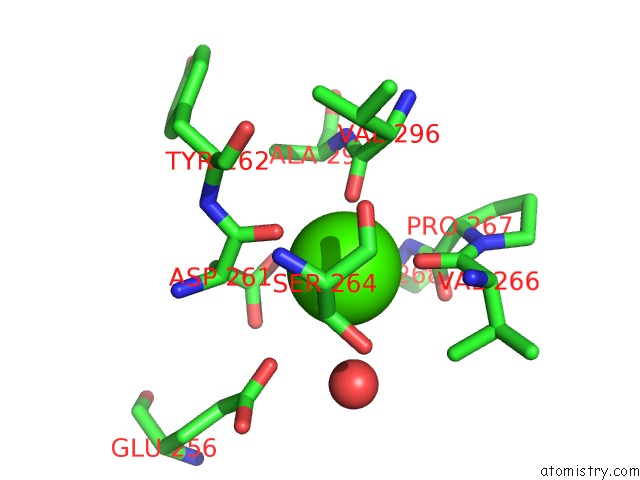
Mono view
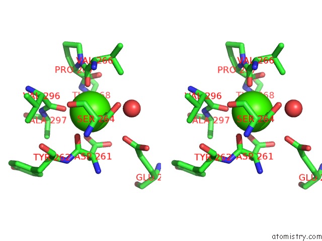
Stereo pair view
Mono view
Stereo pair view
A full contact list of Calcium with other atoms in the Ca binding
site number 1 of Structure of the Multifunctional Paramyxovirus Hemagglutinin- Neuraminidase within 5.0Å range:
|
Reference:
S.Crennell,
T.Takimoto,
A.Portner,
G.Taylor.
Crystal Structure of the Multifunctional Paramyxovirus Hemagglutinin-Neuraminidase Nat.Struct.Biol. V. 7 1068 2000.
ISSN: ISSN 1072-8368
PubMed: 11062565
DOI: 10.1038/81002
Page generated: Thu Jul 11 07:47:38 2024
ISSN: ISSN 1072-8368
PubMed: 11062565
DOI: 10.1038/81002
Last articles
Zn in 9J0NZn in 9J0O
Zn in 9J0P
Zn in 9FJX
Zn in 9EKB
Zn in 9C0F
Zn in 9CAH
Zn in 9CH0
Zn in 9CH3
Zn in 9CH1