Calcium »
PDB 1edm-1evu »
1ee6 »
Calcium in PDB 1ee6: Crystal Structure of Pectate Lyase From Bacillus Sp. Strain Ksm-P15.
Enzymatic activity of Crystal Structure of Pectate Lyase From Bacillus Sp. Strain Ksm-P15.
All present enzymatic activity of Crystal Structure of Pectate Lyase From Bacillus Sp. Strain Ksm-P15.:
4.2.2.2;
4.2.2.2;
Protein crystallography data
The structure of Crystal Structure of Pectate Lyase From Bacillus Sp. Strain Ksm-P15., PDB code: 1ee6
was solved by
M.Akita,
A.Suzuki,
T.Kobayashi,
S.Ito,
T.Yamane,
with X-Ray Crystallography technique. A brief refinement statistics is given in the table below:
| Resolution Low / High (Å) | 10.00 / 2.30 |
| Space group | P 21 21 21 |
| Cell size a, b, c (Å), α, β, γ (°) | 42.900, 60.500, 83.000, 90.00, 90.00, 90.00 |
| R / Rfree (%) | 18.6 / 23.4 |
Calcium Binding Sites:
The binding sites of Calcium atom in the Crystal Structure of Pectate Lyase From Bacillus Sp. Strain Ksm-P15.
(pdb code 1ee6). This binding sites where shown within
5.0 Angstroms radius around Calcium atom.
In total only one binding site of Calcium was determined in the Crystal Structure of Pectate Lyase From Bacillus Sp. Strain Ksm-P15., PDB code: 1ee6:
In total only one binding site of Calcium was determined in the Crystal Structure of Pectate Lyase From Bacillus Sp. Strain Ksm-P15., PDB code: 1ee6:
Calcium binding site 1 out of 1 in 1ee6
Go back to
Calcium binding site 1 out
of 1 in the Crystal Structure of Pectate Lyase From Bacillus Sp. Strain Ksm-P15.
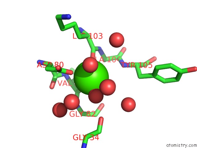
Mono view
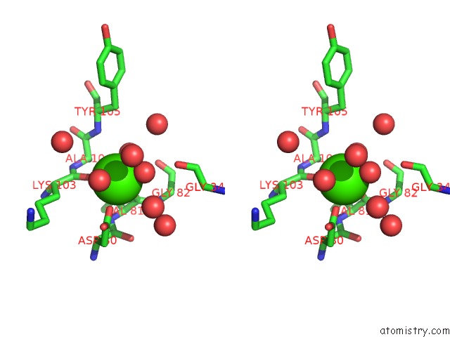
Stereo pair view
Mono view
Stereo pair view
A full contact list of Calcium with other atoms in the Ca binding
site number 1 of Crystal Structure of Pectate Lyase From Bacillus Sp. Strain Ksm-P15. within 5.0Å range:
|
Reference:
M.Akita,
A.Suzuki,
T.Kobayashi,
S.Ito,
T.Yamane.
The First Structure of Pectate Lyase Belonging to Polysaccharide Lyase Family 3. Acta Crystallogr.,Sect.D V. 57 1786 2001.
ISSN: ISSN 0907-4449
PubMed: 11717490
DOI: 10.1107/S0907444900003334
Page generated: Mon Jul 7 14:38:07 2025
ISSN: ISSN 0907-4449
PubMed: 11717490
DOI: 10.1107/S0907444900003334
Last articles
Ca in 7G7FCa in 7G7E
Ca in 7G7C
Ca in 7G7A
Ca in 7G7D
Ca in 7G79
Ca in 7G7B
Ca in 7G78
Ca in 7G74
Ca in 7G77