Calcium »
PDB 1ex0-1fae »
1ezs »
Calcium in PDB 1ezs: Crystal Structure of Ecotin Mutant M84R, W67A, G68A, Y69A, D70A Bound to Rat Anionic Trypsin II
Enzymatic activity of Crystal Structure of Ecotin Mutant M84R, W67A, G68A, Y69A, D70A Bound to Rat Anionic Trypsin II
All present enzymatic activity of Crystal Structure of Ecotin Mutant M84R, W67A, G68A, Y69A, D70A Bound to Rat Anionic Trypsin II:
3.4.21.4;
3.4.21.4;
Protein crystallography data
The structure of Crystal Structure of Ecotin Mutant M84R, W67A, G68A, Y69A, D70A Bound to Rat Anionic Trypsin II, PDB code: 1ezs
was solved by
S.A.Gillmor,
T.Takeuchi,
S.Q.Yang,
C.S.Craik,
R.J.Fletterick,
with X-Ray Crystallography technique. A brief refinement statistics is given in the table below:
| Resolution Low / High (Å) | 25.00 / 2.30 |
| Space group | P 1 |
| Cell size a, b, c (Å), α, β, γ (°) | 45.828, 59.682, 71.044, 103.57, 93.27, 94.62 |
| R / Rfree (%) | 18.8 / 23.5 |
Calcium Binding Sites:
The binding sites of Calcium atom in the Crystal Structure of Ecotin Mutant M84R, W67A, G68A, Y69A, D70A Bound to Rat Anionic Trypsin II
(pdb code 1ezs). This binding sites where shown within
5.0 Angstroms radius around Calcium atom.
In total 2 binding sites of Calcium where determined in the Crystal Structure of Ecotin Mutant M84R, W67A, G68A, Y69A, D70A Bound to Rat Anionic Trypsin II, PDB code: 1ezs:
Jump to Calcium binding site number: 1; 2;
In total 2 binding sites of Calcium where determined in the Crystal Structure of Ecotin Mutant M84R, W67A, G68A, Y69A, D70A Bound to Rat Anionic Trypsin II, PDB code: 1ezs:
Jump to Calcium binding site number: 1; 2;
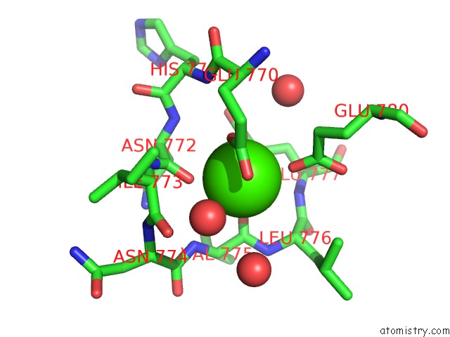
Calcium binding site 1 out of 2 in 1ezs
Go back to
Calcium binding site 1 out
of 2 in the Crystal Structure of Ecotin Mutant M84R, W67A, G68A, Y69A, D70A Bound to Rat Anionic Trypsin II

Mono view

Stereo pair view
Mono view
Stereo pair view
A full contact list of Calcium with other atoms in the Ca binding
site number 1 of Crystal Structure of Ecotin Mutant M84R, W67A, G68A, Y69A, D70A Bound to Rat Anionic Trypsin II within 5.0Å range:
|
Calcium binding site 2 out of 2 in 1ezs
Go back to
Calcium binding site 2 out
of 2 in the Crystal Structure of Ecotin Mutant M84R, W67A, G68A, Y69A, D70A Bound to Rat Anionic Trypsin II
Mono view

Stereo pair view
Mono view
Stereo pair view
A full contact list of Calcium with other atoms in the Ca binding
site number 2 of Crystal Structure of Ecotin Mutant M84R, W67A, G68A, Y69A, D70A Bound to Rat Anionic Trypsin II within 5.0Å range:
|
Reference:
S.A.Gillmor,
T.Takeuchi,
S.Q.Yang,
C.S.Craik,
R.J.Fletterick.
Compromise and Accommodation in Ecotin, A Dimeric Macromolecular Inhibitor of Serine Proteases. J.Mol.Biol. V. 299 993 2000.
ISSN: ISSN 0022-2836
PubMed: 10843853
DOI: 10.1006/JMBI.2000.3812
Page generated: Thu Jul 11 07:59:21 2024
ISSN: ISSN 0022-2836
PubMed: 10843853
DOI: 10.1006/JMBI.2000.3812
Last articles
Zn in 9MJ5Zn in 9HNW
Zn in 9G0L
Zn in 9FNE
Zn in 9DZN
Zn in 9E0I
Zn in 9D32
Zn in 9DAK
Zn in 8ZXC
Zn in 8ZUF