Calcium »
PDB 1ex0-1fae »
1f0u »
Calcium in PDB 1f0u: Bovine Trypsin Complexed with RPR128515
Enzymatic activity of Bovine Trypsin Complexed with RPR128515
All present enzymatic activity of Bovine Trypsin Complexed with RPR128515:
3.4.21.4;
3.4.21.4;
Protein crystallography data
The structure of Bovine Trypsin Complexed with RPR128515, PDB code: 1f0u
was solved by
S.Maignan,
J.P.Guilloteau,
S.Pouzieux,
Y.M.Choi-Sledeski,
M.R.Becker,
S.I.Klein,
W.R.Ewing,
H.W.Pauls,
A.P.Spada,
V.Mikol,
with X-Ray Crystallography technique. A brief refinement statistics is given in the table below:
| Resolution Low / High (Å) | 15.00 / 1.90 |
| Space group | P 21 21 21 |
| Cell size a, b, c (Å), α, β, γ (°) | 63.730, 63.890, 69.170, 90.00, 90.00, 90.00 |
| R / Rfree (%) | 17.2 / n/a |
Calcium Binding Sites:
The binding sites of Calcium atom in the Bovine Trypsin Complexed with RPR128515
(pdb code 1f0u). This binding sites where shown within
5.0 Angstroms radius around Calcium atom.
In total only one binding site of Calcium was determined in the Bovine Trypsin Complexed with RPR128515, PDB code: 1f0u:
In total only one binding site of Calcium was determined in the Bovine Trypsin Complexed with RPR128515, PDB code: 1f0u:
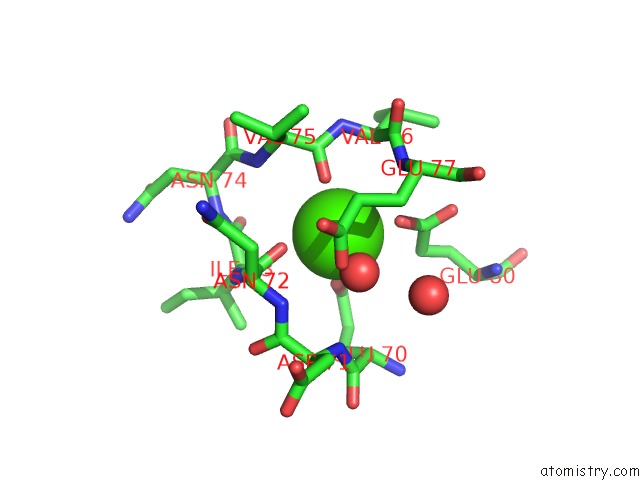
Calcium binding site 1 out of 1 in 1f0u
Go back to
Calcium binding site 1 out
of 1 in the Bovine Trypsin Complexed with RPR128515
Mono view

Stereo pair view
Mono view
Stereo pair view
A full contact list of Calcium with other atoms in the Ca binding
site number 1 of Bovine Trypsin Complexed with RPR128515 within 5.0Å range:
|
Reference:
S.Maignan,
J.P.Guilloteau,
S.Pouzieux,
Y.M.Choi-Sledeski,
M.R.Becker,
S.I.Klein,
W.R.Ewing,
H.W.Pauls,
A.P.Spada,
V.Mikol.
Crystal Structures of Human Factor Xa Complexed with Potent Inhibitors. J.Med.Chem. V. 43 3226 2000.
ISSN: ISSN 0022-2623
PubMed: 10966741
DOI: 10.1021/JM000940U
Page generated: Mon Jul 7 14:45:13 2025
ISSN: ISSN 0022-2623
PubMed: 10966741
DOI: 10.1021/JM000940U
Last articles
Fe in 2YXOFe in 2YRS
Fe in 2YXC
Fe in 2YNM
Fe in 2YVJ
Fe in 2YP1
Fe in 2YU2
Fe in 2YU1
Fe in 2YQB
Fe in 2YOO