Calcium »
PDB 1ex0-1fae »
1f9o »
Calcium in PDB 1f9o: Crystal Structure of the Cellulase CEL48F From C. Cellulolyticum with the Thiooligosaccharide Inhibitor Pips-IG3
Enzymatic activity of Crystal Structure of the Cellulase CEL48F From C. Cellulolyticum with the Thiooligosaccharide Inhibitor Pips-IG3
All present enzymatic activity of Crystal Structure of the Cellulase CEL48F From C. Cellulolyticum with the Thiooligosaccharide Inhibitor Pips-IG3:
3.2.1.4;
3.2.1.4;
Protein crystallography data
The structure of Crystal Structure of the Cellulase CEL48F From C. Cellulolyticum with the Thiooligosaccharide Inhibitor Pips-IG3, PDB code: 1f9o
was solved by
G.Parsiegla,
C.Reverbel-Leroy,
C.Tardif,
J.P.Belaich,
H.Driguez,
R.Haser,
with X-Ray Crystallography technique. A brief refinement statistics is given in the table below:
| Resolution Low / High (Å) | 19.96 / 2.50 |
| Space group | P 21 21 21 |
| Cell size a, b, c (Å), α, β, γ (°) | 61.600, 84.570, 121.820, 90.00, 90.00, 90.00 |
| R / Rfree (%) | 17.9 / 24.3 |
Other elements in 1f9o:
The structure of Crystal Structure of the Cellulase CEL48F From C. Cellulolyticum with the Thiooligosaccharide Inhibitor Pips-IG3 also contains other interesting chemical elements:
| Iodine | (I) | 2 atoms |
Calcium Binding Sites:
The binding sites of Calcium atom in the Crystal Structure of the Cellulase CEL48F From C. Cellulolyticum with the Thiooligosaccharide Inhibitor Pips-IG3
(pdb code 1f9o). This binding sites where shown within
5.0 Angstroms radius around Calcium atom.
In total only one binding site of Calcium was determined in the Crystal Structure of the Cellulase CEL48F From C. Cellulolyticum with the Thiooligosaccharide Inhibitor Pips-IG3, PDB code: 1f9o:
In total only one binding site of Calcium was determined in the Crystal Structure of the Cellulase CEL48F From C. Cellulolyticum with the Thiooligosaccharide Inhibitor Pips-IG3, PDB code: 1f9o:
Calcium binding site 1 out of 1 in 1f9o
Go back to
Calcium binding site 1 out
of 1 in the Crystal Structure of the Cellulase CEL48F From C. Cellulolyticum with the Thiooligosaccharide Inhibitor Pips-IG3
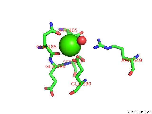
Mono view
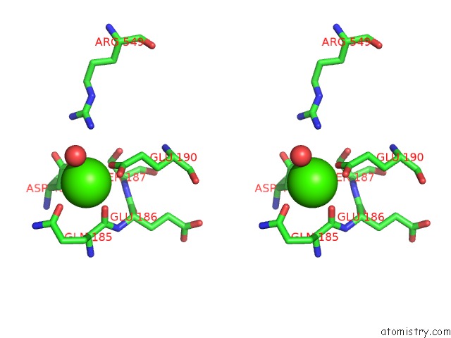
Stereo pair view
Mono view
Stereo pair view
A full contact list of Calcium with other atoms in the Ca binding
site number 1 of Crystal Structure of the Cellulase CEL48F From C. Cellulolyticum with the Thiooligosaccharide Inhibitor Pips-IG3 within 5.0Å range:
|
Reference:
G.Parsiegla,
C.Reverbel-Leroy,
C.Tardif,
J.P.Belaich,
H.Driguez,
R.Haser.
Crystal Structures of the Cellulase CEL48F in Complex with Inhibitors and Substrates Give Insights Into Its Processive Action Biochemistry V. 39 11238 2000.
ISSN: ISSN 0006-2960
PubMed: 10985769
DOI: 10.1021/BI001139P
Page generated: Thu Jul 11 08:04:36 2024
ISSN: ISSN 0006-2960
PubMed: 10985769
DOI: 10.1021/BI001139P
Last articles
Zn in 9MJ5Zn in 9HNW
Zn in 9G0L
Zn in 9FNE
Zn in 9DZN
Zn in 9E0I
Zn in 9D32
Zn in 9DAK
Zn in 8ZXC
Zn in 8ZUF