Calcium »
PDB 1fak-1fn6 »
1fid »
Calcium in PDB 1fid: Structure of Human Gamma Fibrinogen 30 Kd Carboxyl Terminal Fragment
Protein crystallography data
The structure of Structure of Human Gamma Fibrinogen 30 Kd Carboxyl Terminal Fragment, PDB code: 1fid
was solved by
V.C.Yee,
D.C.Teller,
with X-Ray Crystallography technique. A brief refinement statistics is given in the table below:
| Resolution Low / High (Å) | 10.00 / 2.10 |
| Space group | P 21 21 2 |
| Cell size a, b, c (Å), α, β, γ (°) | 84.360, 62.560, 49.620, 90.00, 90.00, 90.00 |
| R / Rfree (%) | 17 / 22.6 |
Calcium Binding Sites:
The binding sites of Calcium atom in the Structure of Human Gamma Fibrinogen 30 Kd Carboxyl Terminal Fragment
(pdb code 1fid). This binding sites where shown within
5.0 Angstroms radius around Calcium atom.
In total only one binding site of Calcium was determined in the Structure of Human Gamma Fibrinogen 30 Kd Carboxyl Terminal Fragment, PDB code: 1fid:
In total only one binding site of Calcium was determined in the Structure of Human Gamma Fibrinogen 30 Kd Carboxyl Terminal Fragment, PDB code: 1fid:
Calcium binding site 1 out of 1 in 1fid
Go back to
Calcium binding site 1 out
of 1 in the Structure of Human Gamma Fibrinogen 30 Kd Carboxyl Terminal Fragment
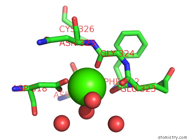
Mono view
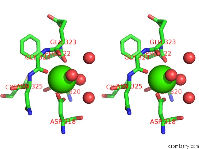
Stereo pair view
Mono view
Stereo pair view
A full contact list of Calcium with other atoms in the Ca binding
site number 1 of Structure of Human Gamma Fibrinogen 30 Kd Carboxyl Terminal Fragment within 5.0Å range:
|
Reference:
V.C.Yee,
K.P.Pratt,
H.C.Cote,
I.L.Trong,
D.W.Chung,
E.W.Davie,
R.E.Stenkamp,
D.C.Teller.
Crystal Structure of A 30 kDa C-Terminal Fragment From the Gamma Chain of Human Fibrinogen. Structure V. 5 125 1997.
ISSN: ISSN 0969-2126
PubMed: 9016719
DOI: 10.1016/S0969-2126(97)00171-8
Page generated: Thu Jul 11 08:09:11 2024
ISSN: ISSN 0969-2126
PubMed: 9016719
DOI: 10.1016/S0969-2126(97)00171-8
Last articles
Zn in 9J0NZn in 9J0O
Zn in 9J0P
Zn in 9FJX
Zn in 9EKB
Zn in 9C0F
Zn in 9CAH
Zn in 9CH0
Zn in 9CH3
Zn in 9CH1