Calcium »
PDB 1fak-1fn6 »
1fmi »
Calcium in PDB 1fmi: Crystal Structure of Human Class I ALPHA1,2-Mannosidase
Enzymatic activity of Crystal Structure of Human Class I ALPHA1,2-Mannosidase
All present enzymatic activity of Crystal Structure of Human Class I ALPHA1,2-Mannosidase:
3.2.1.113;
3.2.1.113;
Protein crystallography data
The structure of Crystal Structure of Human Class I ALPHA1,2-Mannosidase, PDB code: 1fmi
was solved by
F.Vallee,
K.Karaveg,
A.Herscovics,
K.W.Moremen,
P.L.Howell,
with X-Ray Crystallography technique. A brief refinement statistics is given in the table below:
| Resolution Low / High (Å) | 28.52 / 1.90 |
| Space group | P 31 2 1 |
| Cell size a, b, c (Å), α, β, γ (°) | 95.829, 95.829, 136.853, 90.00, 90.00, 120.00 |
| R / Rfree (%) | 22.2 / 25 |
Calcium Binding Sites:
The binding sites of Calcium atom in the Crystal Structure of Human Class I ALPHA1,2-Mannosidase
(pdb code 1fmi). This binding sites where shown within
5.0 Angstroms radius around Calcium atom.
In total only one binding site of Calcium was determined in the Crystal Structure of Human Class I ALPHA1,2-Mannosidase, PDB code: 1fmi:
In total only one binding site of Calcium was determined in the Crystal Structure of Human Class I ALPHA1,2-Mannosidase, PDB code: 1fmi:
Calcium binding site 1 out of 1 in 1fmi
Go back to
Calcium binding site 1 out
of 1 in the Crystal Structure of Human Class I ALPHA1,2-Mannosidase
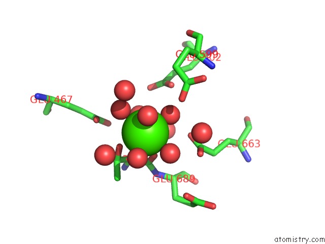
Mono view
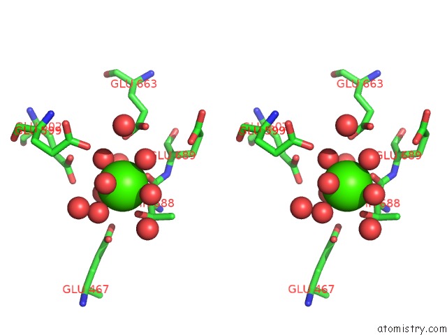
Stereo pair view
Mono view
Stereo pair view
A full contact list of Calcium with other atoms in the Ca binding
site number 1 of Crystal Structure of Human Class I ALPHA1,2-Mannosidase within 5.0Å range:
|
Reference:
F.Vallee,
K.Karaveg,
A.Herscovics,
K.W.Moremen,
P.L.Howell.
Structural Basis For Catalysis and Inhibition of N-Glycan Processing Class I Alpha 1,2-Mannosidases. J.Biol.Chem. V. 275 41287 2000.
ISSN: ISSN 0021-9258
PubMed: 10995765
DOI: 10.1074/JBC.M006927200
Page generated: Thu Jul 11 08:14:51 2024
ISSN: ISSN 0021-9258
PubMed: 10995765
DOI: 10.1074/JBC.M006927200
Last articles
Zn in 9J0NZn in 9J0O
Zn in 9J0P
Zn in 9FJX
Zn in 9EKB
Zn in 9C0F
Zn in 9CAH
Zn in 9CH0
Zn in 9CH3
Zn in 9CH1