Calcium »
PDB 1fak-1fn6 »
1fml »
Calcium in PDB 1fml: Crystal Structure of Retinol Dehydratase in A Complex with Retinol and Pap
Protein crystallography data
The structure of Crystal Structure of Retinol Dehydratase in A Complex with Retinol and Pap, PDB code: 1fml
was solved by
S.Pakhomova,
M.Kobayashi,
J.Buck,
M.E.Newcomer,
with X-Ray Crystallography technique. A brief refinement statistics is given in the table below:
| Resolution Low / High (Å) | 38.45 / 2.75 |
| Space group | P 1 21 1 |
| Cell size a, b, c (Å), α, β, γ (°) | 82.053, 66.612, 84.904, 90.00, 111.29, 90.00 |
| R / Rfree (%) | 22.2 / 27.3 |
Calcium Binding Sites:
The binding sites of Calcium atom in the Crystal Structure of Retinol Dehydratase in A Complex with Retinol and Pap
(pdb code 1fml). This binding sites where shown within
5.0 Angstroms radius around Calcium atom.
In total 2 binding sites of Calcium where determined in the Crystal Structure of Retinol Dehydratase in A Complex with Retinol and Pap, PDB code: 1fml:
Jump to Calcium binding site number: 1; 2;
In total 2 binding sites of Calcium where determined in the Crystal Structure of Retinol Dehydratase in A Complex with Retinol and Pap, PDB code: 1fml:
Jump to Calcium binding site number: 1; 2;
Calcium binding site 1 out of 2 in 1fml
Go back to
Calcium binding site 1 out
of 2 in the Crystal Structure of Retinol Dehydratase in A Complex with Retinol and Pap

Mono view
Stereo pair view
Mono view
Stereo pair view
A full contact list of Calcium with other atoms in the Ca binding
site number 1 of Crystal Structure of Retinol Dehydratase in A Complex with Retinol and Pap within 5.0Å range:
|
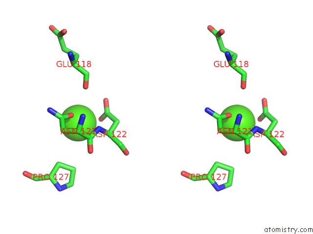
Calcium binding site 2 out of 2 in 1fml
Go back to
Calcium binding site 2 out
of 2 in the Crystal Structure of Retinol Dehydratase in A Complex with Retinol and Pap

Mono view

Stereo pair view
Mono view
Stereo pair view
A full contact list of Calcium with other atoms in the Ca binding
site number 2 of Crystal Structure of Retinol Dehydratase in A Complex with Retinol and Pap within 5.0Å range:
|
Reference:
S.Pakhomova,
M.Kobayashi,
J.Buck,
M.E.Newcomer.
A Helical Lid Converts A Sulfotransferase to A Dehydratase. Nat.Struct.Biol. V. 8 447 2001.
ISSN: ISSN 1072-8368
PubMed: 11323722
DOI: 10.1038/87617
Page generated: Mon Jul 7 14:55:47 2025
ISSN: ISSN 1072-8368
PubMed: 11323722
DOI: 10.1038/87617
Last articles
F in 7M8VF in 7MCK
F in 7MAZ
F in 7MBO
F in 7MCE
F in 7MB2
F in 7MB1
F in 7MAX
F in 7M8R
F in 7M9R