Calcium »
PDB 1fn7-1fzc »
1fo4 »
Calcium in PDB 1fo4: Crystal Structure of Xanthine Dehydrogenase Isolated From Bovine Milk
Enzymatic activity of Crystal Structure of Xanthine Dehydrogenase Isolated From Bovine Milk
All present enzymatic activity of Crystal Structure of Xanthine Dehydrogenase Isolated From Bovine Milk:
1.1.1.204;
1.1.1.204;
Protein crystallography data
The structure of Crystal Structure of Xanthine Dehydrogenase Isolated From Bovine Milk, PDB code: 1fo4
was solved by
C.Enroth,
B.T.Eger,
K.Okamoto,
T.Nishino,
T.Nishino,
E.F.Pai,
with X-Ray Crystallography technique. A brief refinement statistics is given in the table below:
| Resolution Low / High (Å) | 25.00 / 2.10 |
| Space group | C 1 2 1 |
| Cell size a, b, c (Å), α, β, γ (°) | 169.451, 124.493, 148.327, 90.00, 90.94, 90.00 |
| R / Rfree (%) | 19.8 / 23.8 |
Other elements in 1fo4:
The structure of Crystal Structure of Xanthine Dehydrogenase Isolated From Bovine Milk also contains other interesting chemical elements:
| Molybdenum | (Mo) | 2 atoms |
| Iron | (Fe) | 8 atoms |
Calcium Binding Sites:
The binding sites of Calcium atom in the Crystal Structure of Xanthine Dehydrogenase Isolated From Bovine Milk
(pdb code 1fo4). This binding sites where shown within
5.0 Angstroms radius around Calcium atom.
In total 2 binding sites of Calcium where determined in the Crystal Structure of Xanthine Dehydrogenase Isolated From Bovine Milk, PDB code: 1fo4:
Jump to Calcium binding site number: 1; 2;
In total 2 binding sites of Calcium where determined in the Crystal Structure of Xanthine Dehydrogenase Isolated From Bovine Milk, PDB code: 1fo4:
Jump to Calcium binding site number: 1; 2;
Calcium binding site 1 out of 2 in 1fo4
Go back to
Calcium binding site 1 out
of 2 in the Crystal Structure of Xanthine Dehydrogenase Isolated From Bovine Milk
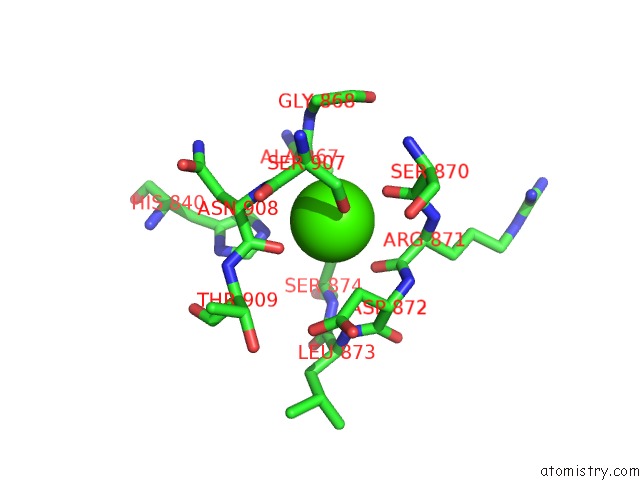
Mono view
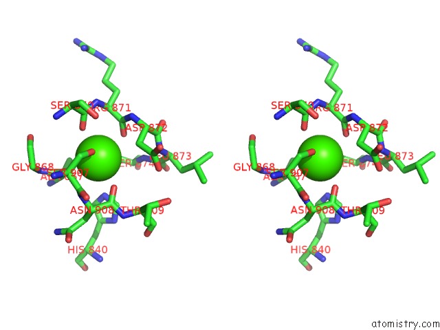
Stereo pair view
Mono view
Stereo pair view
A full contact list of Calcium with other atoms in the Ca binding
site number 1 of Crystal Structure of Xanthine Dehydrogenase Isolated From Bovine Milk within 5.0Å range:
|
Calcium binding site 2 out of 2 in 1fo4
Go back to
Calcium binding site 2 out
of 2 in the Crystal Structure of Xanthine Dehydrogenase Isolated From Bovine Milk
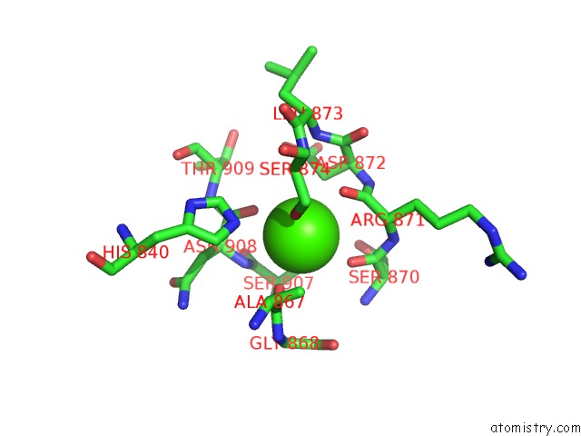
Mono view
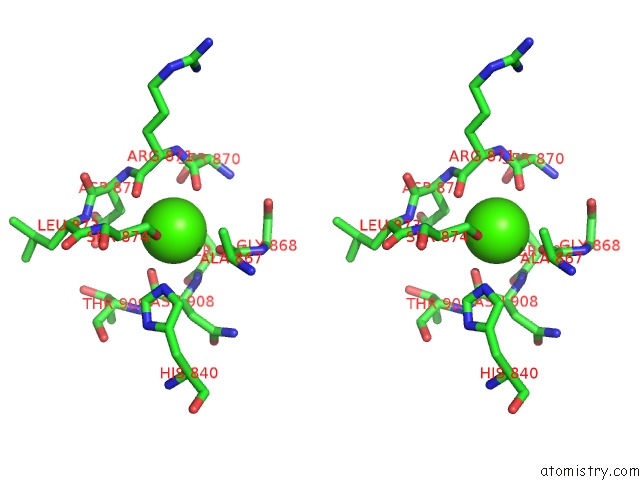
Stereo pair view
Mono view
Stereo pair view
A full contact list of Calcium with other atoms in the Ca binding
site number 2 of Crystal Structure of Xanthine Dehydrogenase Isolated From Bovine Milk within 5.0Å range:
|
Reference:
C.Enroth,
B.T.Eger,
K.Okamoto,
T.Nishino,
T.Nishino,
E.F.Pai.
Crystal Structures of Bovine Milk Xanthine Dehydrogenase and Xanthine Oxidase: Structure-Based Mechanism of Conversion. Proc.Natl.Acad.Sci.Usa V. 97 10723 2000.
ISSN: ISSN 0027-8424
PubMed: 11005854
DOI: 10.1073/PNAS.97.20.10723
Page generated: Thu Jul 11 08:16:26 2024
ISSN: ISSN 0027-8424
PubMed: 11005854
DOI: 10.1073/PNAS.97.20.10723
Last articles
Zn in 9J0NZn in 9J0O
Zn in 9J0P
Zn in 9FJX
Zn in 9EKB
Zn in 9C0F
Zn in 9CAH
Zn in 9CH0
Zn in 9CH3
Zn in 9CH1